. 2006 Nov 15;177(10):6593-7.
doi: 10.4049/jimmunol.177.10.6593.

Cutting edge: inherent and acquired resistance to radiation-induced apoptosis in B cells: a pivotal role for STAT3

Affiliations

Cutting edge: inherent and acquired resistance to radiation-induced apoptosis in B cells: a pivotal role for STAT3

Dennis C Otero et al. J Immunol. .

Abstract

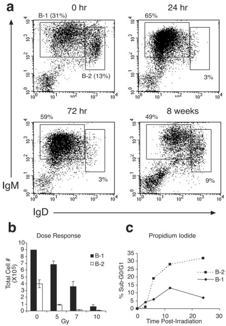
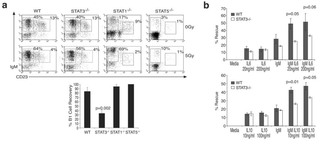
Radiation-induced apoptosis (RiA) is used therapeutically for tumor cell ablation as well as a tool to characterize hemopoietic cell lineages. We report that the peritoneal B-1 B cell subset is selectively resistant to RiA. Inherent radioresistance is not shared by splenic B-2 or B-1 cells. However, it is conferred upon B-2 cells by BCR crosslinking in the presence of IL-6 or IL-10. In vivo experiments with gene-targeted mice confirm that IL-6 and, to a lesser extent, IL-10 are the relevant stimuli that combine with BCR ligands to promote B-1 cell radioresistance. STAT3 promotes cell survival in response to selected growth factors, and is activated by combined BCR crosslinking and IL-6 (IL-10). Importantly, STAT3(-/-) B-1 cells become susceptible to irradiation, indicating that STAT3 activation by the BCR in the presence of IL costimuli account for the inherent radioresistance of peritoneal B-1 B cells.


Conflict of interest statement

Disclosures

The authors have no financial conflict of interest.

Figures

FIGURE 1
FIGURE 1
Murine B-1 cells are radioresistant. a, Flow cytometry profiles of peritoneal B-1 (IgMhighIgDlow) and B-2 (IgMlowIgDhigh) cells at indicated time points postirradiation (5 Gy) (n = 6 mice). b, B cell recovery from PeC 48 h after indicated exposure (n = 3 per group). c, Kinetics of in vitro cell death determined by PI staining after 2 Gy exposure; representative of more than three experiments.
FIGURE 2
FIGURE 2
Cytokines and cell activation contribute to B cell radioresistance. a, Recovery of splenic B-1 cells from VH12 transgenic mice 48 h postirradiation (5 Gy, n = 3 mice). b, Rescue of B-2 cells from RiA. Purified CFSE-labeled splenic B-2 cells were left untreated or stimulated with anti-IgM (10 µg/ml) and immediately injected i.p. or i.v. Recipient mice were irradiated (5 Gy) and transferred B cells from PeC or spleen were enumerated 24 h postirradiation. Graph represents percent cell recovery from irradiated mice compared with non-irradiated mice (n = 3 mice per group). c, In vitro rescue of B cells from RiA. Purified splenic B cells were irradiated (2 Gy) following an 18-h incubation with anti-IgM and/or IL-6. Apoptosis was measured by PI (sub-G0-G1) staining 24 h postirradiation; representative of four experiments. d, Expression levels of IL-6 and IL-10 receptor on ex vivo and stimulated (24 h) B cells; LPS (1 µg/ml), anti-CD40 (10 µg/ml), and anti-IgM (1 µg/ml). e, B cell recovery from the PeC of IL-6−/− and IL-10−/− mice. Relative numbers of B-1 (CD23negIgMhigh) and B-2 (CD23posIgMlow) cells were analyzed before and after (48 h) irradiation (5 Gy). Graph represents percent cell recovery from the PeC of irradiated mice compared with non-irradiated mice (average ± SEM of at least three mice per group).
FIGURE 3
FIGURE 3
B-1 cell radioresistance requires STAT3. a, B cell recovery from the PeC of STAT3−/−, STAT1−/−, and STAT5a/b−/− mice. Relative numbers of B-1 and B-2 cells were determined before and after (48 h) irradiation (5 Gy). Graph represents percent cell recovery from the PeC of irradiated mice compared with non-irradiated mice (average ± SEM of at ≥3 mice per group). b, Impaired in vitro rescue of STAT3−/− B cells from RiA. Purified splenic B cells from WT and STAT-3−/− mice were cultured (18 h) in the presence of anti-IgM and/or IL-6 (top) or anti-IgM/IL-10 (bottom), irradiated (2 Gy), and apoptosis measured by PI (sub-G0-G1) staining 24 h postirradiation; representative of three experiments (mean ± SEM).

References

    1. Berland R, Wortis HH. Origins and functions of B-1 cells with notes on the role of CD5. Annu. Rev. Immunol. 2002;20:253–300. - PubMed
    1. Montecino-Rodriguez E, Leathers H, Dorshkind K. Identification of a B-1 B cell-specified progenitor. Nat. Immunol. 2006;7:293–301. - PubMed
    1. Hardy RR, Hayakawa K. A Developmental Switch in B Lymphopoiesis. PNAS. 1991;88:11550–11554. - PMC - PubMed
    1. Hayakawa K, Asano M, Shinton SA, Gui M, Allman D, Stewart CL, Silver J, Hardy RR. Positive selection of natural autoreactive B cells. Science. 1999;285:113–116. - PubMed
    1. Arnold LW, Pennell CA, McCray SK, Clarke SH. Development of B-1 cells: segregation of phosphatidyl choline-specific B cells to the B-1 population occurs after immunoglobulin gene expression. J. Exp. Med. 1994;179:1585–1595. - PMC - PubMed

Publication types

MeSH terms