. 2006 Nov 15;25(22):5396-404.
doi: 10.1038/sj.emboj.7601409. Epub 2006 Nov 2.

Ferroportin-mediated mobilization of ferritin iron precedes ferritin degradation by the proteasome

Affiliations

Ferroportin-mediated mobilization of ferritin iron precedes ferritin degradation by the proteasome

Ivana De Domenico et al. EMBO J. .

Abstract

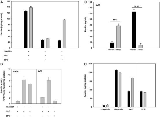
Ferritin is a cytosolic molecule comprised of subunits that self-assemble into a nanocage capable of containing up to 4500 iron atoms. Iron stored within ferritin can be mobilized for use within cells or exported from cells. Expression of ferroportin (Fpn) results in export of cytosolic iron and ferritin degradation. Fpn-mediated iron loss from ferritin occurs in the cytosol and precedes ferritin degradation by the proteasome. Depletion of ferritin iron induces the monoubiquitination of ferritin subunits. Ubiquitination is not required for iron release but is required for disassembly of ferritin nanocages, which is followed by degradation of ferritin by the proteasome. Specific mammalian machinery is not required to extract iron from ferritin. Iron can be removed from ferritin when ferritin is expressed in Saccharomyces cerevisiae, which does not have endogenous ferritin. Expressed ferritin is monoubiquitinated and degraded by the proteasome. Exposure of ubiquitination defective mammalian cells to the iron chelator desferrioxamine leads to degradation of ferritin in the lysosome, which can be prevented by inhibitors of autophagy. Thus, ferritin degradation can occur through two different mechanisms.


Figures

Figure 1
Figure 1
Fpn-mediated decrease in ferritin is not prevented by chloroquine or leupeptin. (A) HEK293T-Fpn cells were incubated with FAC (10 μM Fe) for 24 h followed by incubation for 6 h in the absence or presence of 10 μM Ponasterone A. Cells were then incubated with and without 100 μM chloroquine or 10 μM leupeptin for 10 h and harvested. The ferritin content was determined by ELISA. Induction of Fpn resulted in decreased ferritin levels and this was not prevented by treatment with chloroquine or leupeptin. The data are presented as the standard deviation from three different experiments. (B) Cells treated as in (A) were incubated in the presence of 1 μg/ml EGF for 2 h. Cells were fixed and processed for immunofluorescence using mouse anti-EGF receptor and Alexa 594 conjugated goat anti-mouse IgG. Chloroquine and leupeptin effectively inhibit degradation of EGF receptor.
Figure 2
Figure 2
Fpn-mediated decrease in ferritin results from degradation by the proteasome. (A) HEK293T-Fpn cells were incubated with FAC (10 μM Fe) for 24 h followed by incubation for 6 h in the absence or presence of 10 μM Ponasterone A. Cells were then treated with or without 10 μM MG132 or 10 μM lactacystin in the presence of Ponasterone A for 10 h and harvested. The ferritin content was determined by ELISA. Error bars represent the standard deviation from three different experiments in duplicate. (B) Samples treated as in (A) were immunoprecipitated using anti-ferritin antibodies and the immunoprecipitate examined for the presence of ferritin or ubiquitin by Western blot analysis. The arrows indicate the migration of H and L chains. (C) Cells were incubated with 1.0 × 10⁻⁷ M Tf(59Fe)2 for 24 h followed by incubation for 6 h in the absence or presence of 10 μM Ponasterone A. Cells were then treated with or without 10 μM MG132 in the presence of Ponasterone A for 10 h and harvested and the amount of 59Fe in immunoprecipitated ferritin was determined. (D) Cells were treated as described in (A) and cell extracts were applied to size exclusion chromatography and the ferritin content, in selected fractions, was determined by ELISA. Error bars represent the standard deviation from three different experiments in duplicate. (E) Cells were treated as described in (C), ferritin was then immunoprecipitated, eluted using 100 mM glycine, pH 2.5 and measured by ELISA. The amount of ferritin-associated 59Fe was measured and the specific activity of ferritin determined. Error bars represent the standard deviation from three different experiments in duplicate. (F) Samples were treated as in (D), and ferritin was immunoprecipitated and analyzed by Western blot analysis using an antibody to ubiquitin.
Figure 3
Figure 3
Ubiquitination is required for Fpn-mediated ferritin degradation. (A) FM3A (black) and ts85 (grey) cells were transiently transfected with a plasmid containing CMV-regulated Fpn-GFP and incubated in the presence of FAC (10 μM Fe) and 0.5 μM hepcidin for 24 h. Hepcidin was either maintained (+) or removed (−) to allow Fpn-GFP localization at the plasma membrane. Cells were maintained at the permissive temperature (33°C) or moved to the restrictive temperature (39°C) and incubated for 6 h. Cells were harvested and ferritin content determined by ELISA. Error bars represent the standard deviation from three different experiments in duplicate. (B) FM3A and ts85 cells were treated as in (A) but FAC was replaced with 1.0 × 10⁻⁷ M Tf(59Fe)2. Ferritin was immunoprecipitated and the specific activity of 59Fe-ferritin determined. Error bars represent the standard deviation from three different experiments in duplicate. (C) ts85 cells were treated as in (B) and cell extracts applied to size exclusion chromatography. Ferritin content in selected fractions was measured by ELISA. Black bars represent assembled ferritin (>400 kDa), gray bars represent monomeric (<50 kDa). (D) FM3A and ts85 cells were treated as in (A) but cells that had been incubated at the restrictive temperature (39°C) were then returned to the permissive temperature (33°C) and incubated in the presence of cycloheximide (75 μg/ml) and 10 μM FAC for 1 h. Cells were then harvested and ferritin levels determined by ELISA. Error bars represent the standard deviation from three different experiments in duplicate.
Figure 4
Figure 4
DFO leads to lysosomal degradation of ferritin. FM3A and ts85 cells were incubated in the presence of FAC (10 μM Fe) for 12 h. (A) Cells were then incubated at the restrictive temperature (39°C) for 6 h in the presence or absence of 100 μM DFO, with or without 100 μM chloroquine or 10 μM MG132. Cells were then harvested and ferritin content determined by ELISA. Error bars represent the standard deviation from three different experiments in duplicate. (B) Cells treated as in (A) were incubated in the presence or absence of 5 mM 3-methyladenine.
Figure 5
Figure 5
Human ferritin expressed in S. cerevisiae is degraded by the proteasome. Strains of wild type (Wt), Δccc1, erg6-2 cells were transformed with plasmids pGAL, pGAL-L-ferritin, pGAL-H-ferritin and pGAL-H+L-ferritin. (A) Δccc1/pGAL-H+L cells were transformed with a plasmid containing a methionine regulated CCC1 (pMET3CCC1). Cells were grown in medium with galactose for 20 h, washed and incubated in galactose (•), glucose with 10 × methionine (▵), glucose (○) or glucose without methionine (▾) for 10 h. Cells were then harvested and ferritin levels determined by ELISA. Error bars represent the standard deviation from three different experiments in duplicate. The absence of methionine leads to expression of the vacuolar iron transporter Ccc1p. (B) erg6-2 and erg6-2 pGAL-H+L strains were grown in medium with galactose and 250 μM FeSO4 for 24 h. Cells were then washed and incubated in galactose or glucose in the absence or presence of 50 μM MG132 for 7 h. (C) Cells were then harvested, ferritin levels determined by ELISA and immunoprecipitated using anti-ferritin antibodies and the immunoprecipitate examined for the presence of ubiquitin by Western analysis. Cells with the erg6-2 allele permit the entry of MG132 (Lee and Goldberg, 1996).

References

    1. Bridges KR (1987) Ascorbic acid inhibits lysosomal autophagy of ferritin. J Biol Chem 262: 14773–14778 - PubMed
    1. Bridges KR, Hoffman KE (1986) The effects of ascorbic acid on the intracellular metabolism of iron and ferritin. J Biol Chem 261: 14273–14277 - PubMed
    1. Crosby WH (1966) Mucosal block. An evaluation of concepts relating to control of iron absorption. Semin Hematol 3: 299–313 - PubMed
    1. Erhardt JG, Estes JE, Pfeiffer CM, Biesalski HK, Craft NE (2004) Combined measurement of ferritin, soluble transferrin receptor, retinol binding protein, and C-reactive protein by an inexpensive, sensitive, and simple sandwich enzyme-linked immunosorbent assay technique. J Nutr 134: 3127–3132 - PubMed
    1. Finley D, Ciechanover A, Varshavsky A (1984) Thermolability of ubiquitin-activating enzyme from the mammalian cell cycle mutant ts85. Cell 37: 43–55 - PubMed

Publication types