. 2006 Nov 15;25(22):5270-83.
doi: 10.1038/sj.emboj.7601400. Epub 2006 Nov 2.

ADAMTS1 mediates the release of antiangiogenic polypeptides from TSP1 and 2

Affiliations

ADAMTS1 mediates the release of antiangiogenic polypeptides from TSP1 and 2

Nathan V Lee et al. EMBO J. .

Abstract

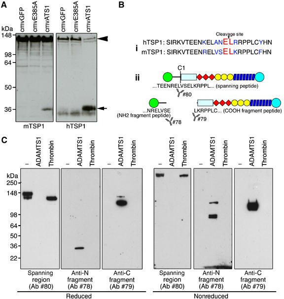
Matrix metalloproteases regulate both physiological and pathological events by processing matrix proteins and growth factors. ADAMTS1 in particular is required for normal ovulation and renal function and has been shown to modulate angiogenesis. Here we report that TSP1 and 2 are substrates of ADAMTS1. Using a combination of mass spectrometry and Edman degradation, we mapped the cleavage sites and characterized the biological relevance of these processing events. ADAMTS1 cleavage mediates the release of polypeptides from the trimeric structure of both TSP1 and 2 generating a pool of antiangiogenic fragments from matrix-bound thrombospondin. Using neo-epitope antibodies we confirmed that processing occurs during wound healing of wild-type mice. However, TSP1 proteolysis is decreased or absent in ADAMTS1 null mice; this is associated with delayed wound closure and increased angiogenic response. Finally, TSP1-/- endothelial cells revealed that the antiangiogenic response mediated by ADAMTS1 is greatly dependent on TSP1. These findings have unraveled a mechanistic explanation for the angiostatic functions attributed to ADAMTS1 and demonstrated in vivo processing of TSP1 under situations of tissue repair.


Figures

Figure 1
Figure 1
TSP1 and 2 are cleaved by ADAMTS1 at unique sites. (A) Coomassie stained gel of full-length TSP1 and Laminin incubated with ADAMTS1. Arrows indicate the 110 and 36 kDa cleavage fragments. (B) Western immunoblot of TSP1 incubated with ADAMTS1 or thrombin. (C) Western immunoblots of TSP1 and 2 incubated with ADAMTS1. (D) Western immunoblot of TSP1 incubated with ADAMTS1, catalytically inactive ADAMTS1 (E385A) or a truncated ADAMTS1 form that only harbors type I repeats (TSRs) for indicated times. (E) Western immunoblot of TSP2 incubated with ADAMTS1, catalytically inactive ADAMTS1 (E385A) or a truncated ADAMTS1 form that only harbors type I repeats (TSRs) for indicated times. Open arrow, fragment already present in preparation that is not susceptible to ADAMTS1 cleavage.
Figure 2
Figure 2
ADAMTS1 cleavage of TSP1 and 2 occurs in a dosage-dependent manner. (A, B) Western immunoblots of TSP1 and 2 incubated with ADAMTS1 for 1 h at 37°C at E:S ranging from 1:1 to 1:40. (C, D) Western immunoblots of TSP1 and 2 incubated with ADAMTS1 in pH ranging from 5.0 to 8.5. Arrowheads, intact TSP1 and 2; arrows, TSP1 and 2 cleavage fragments. Tables under each blot indicate densitometric quantification of the bands. Numbers are in percentile of relative intensity in relation to the darkest band in the blot.
Figure 3
Figure 3
TSP1 and 2 are not substrates of ADAMTS4. (A, B), Western immunoblots of TSP1 and 2 incubated with ADAMTS1, ADAMTS4 and vehicle. (C) Western immunoblots of aggrecan incubated with ADAMTS1, ADAMTS4 or vehicle. (D, E) Western immunoblots of TSP1 and 2 incubated with varying amounts of 87 kDa ADAMTS1 and 65 kDa ADAMTS1, as indicated. Arrowheads, intact TSP1 and 2; arrows, TSP1 and 2 cleavage fragments.
Figure 4
Figure 4
Heparin binding domains are important for cleavage. (A) schematic diagram of full-length TSP1 and 2 and deletion mutants. (B (i, ii), Western immunoblots and Coomassie staining of full-length TSP1 and delN-1 or TSP2 and delN-2 digested with ADAMTS1. (C (i, ii)), Western immunoblots and Coomassie staining of full-length TSP1 and truncated mutant NoC-1truncated or TSP2 and NoC-2 digested with ADAMTS1. Open arrows, ADAMTS1 protein. (D (i, ii)) TSP1 and 2 digested with ADAMTS1 in presence of increasing amounts of heparin. Arrowhead, full-length protein; arrows, cleaved fragments.
Figure 5
Figure 5
TSP1 and 2 cleavage sites and schematic representation of resulting fragments. (A) TSP1 is cleaved at one site C1 by ADAMTS1 adjacent to amino-acid sequence LRRPPL (determined by Edman degradation sequencing). (B) TSP2 is cleaved at two sites C1 and C2. Site C1 is adjacent to amino acids LIGGPP. Proteins were visualized with Coomassie. Asterisk in (A) represents ADAMTS1.
Figure 6
Figure 6
N-terminal fragments remain trimerized releasing the monomeric C-terminal fragments. (A) Sequence alignment of TSP1 and 2 including region in proximity of cleavage sites. Boxed region represents pro-collagen domain. Underlined sequence corresponds to the coiled-coil region. Asterisks denote cysteines involved in interchain disulfide bonds. Inset, schematic representation of N-terminal and C-terminal fragments in reducing (R) and nonreducing (NR) conditions. (B, C) Western immunoblots of TSP1 and 2 incubated with ADAMTS1 or vehicle under nonreducing and reducing conditions.
Figure 7
Figure 7
Characterization of TSP1 neo-epitope antibodies. (A) Western immunoblot of murine and human TSP1 incubated with CM from adenovirus infected cells expressing GFP (cmvGFP), inactive ADAMTS1 (cmvE385A) or full-length active ADAMTS1 (cmvATS1). (B (i)) Sequence alignment of human and murine TSP1 residues flanking cleavage site. (ii) Schematic diagram of TSP1 and fragments recognized by neo-epitope antibodies (78 and 79) and antibody to spanning region (80). (C) Western immunoblot of intact TSP1 and TSP1 fragments resulting from processing with either thrombin or ADAMTS1 under reducing and nonreducing conditions. Arrowhead, full-length TSP1; arrow, TSP1 fragment.
Figure 8
Figure 8
TSP1 cleavage by ADAMTS1 occurs during excisional wound healing. (A) (a–f) Immunohistochemical staining of intact TSP1 and TSP1 fragments in 2-day excisional wound serial sections using the spanning region antibody (#80) or neo-epitope antibodies (#78 and #79). (Arrow in a–f, invading front of epidermal cells; C: fibrin clot; E: epidermis; F: hair follicles) (A). (g–l), Immunohistochemical staining of intact TSP1 and fragments within fibrin clot. Arrows in (g and j), intact TSP1; arrows in (h), N-terminal TSP1 fragment. (B) Western immunoblot of TSP1 (intact and fragments) immunoprecipitated with anti-TSP1 antibodies (#78, 79 or 80) and anti-occludin (occ). Western immunoblot was performed using either #78, 79 or 80 (arrowhead: C-terminal TSP1 fragment; arrow: N- terminal TSP1 fragment; asterisk: intact TSP1).
Figure 9
Figure 9
ADAMTS1 null animals exhibits delayed wound healing and increased angiogenesis. (A) Hematoxylin and eosin stained 5-day wound cross-sections of wild-type and ADAMTS1 null animals. (B) Excisional wound opening area quantified over 5 days in wild-type and adamts1 null animals. Difference in open wound area between wild-type and adamts1 null from day 1 onward has a P-value <0.005. (C) Hematoxylin and eosin (a, d) anti-PECAM stained (b, c, e, f) blood vessels within wound cross-sections. (c) and (f) are magnified images of boxed area in (b) and (e), respectively. (D) Quantification of blood vessels in wounds. Bars indicate standard error. Difference in mean vessel count between wild-type and adamts1 null has a P-value <0.005.
Figure 10
Figure 10
TSP1 fragments are released by endothelial cells and suppress proliferation. (A) Western immunoblots of TSP1 (intact and fragments) secreted from cells (CM), in the cell layer (CL), released from the matrix (SUP) or matrix bound (matrix) incubated with vehicle or ADAMTS1 at the indicated amounts. Arrowhead, intact TSP1; asterisk, C-term fragment; arrow, N-term fragment. (B) Elution chromatogram of TSP1 fragments. Elution of proteins was monitored by absorbance (a.u. on left) and conductivity is shown on the right. Fractions containing eluted proteins were separated by SDS–PAGE and visualized by Coomassie staining and Western immunoblotting with anti-TSP1 antibodies. FL, full-length TSP1 protein. (C) [3H]thymidine incorporation in bovine aortic endothelial cells stimulated with GF (FGF-2 and VEGF) and treated with vehicle, full-length TSP1, or purified TSP1 cleavage products (36 and 110 kDa). (D) Phase micrograph of cultured primary mouse lung endothelial cells isolated from wild-type and tsp1 null animals at passage 3. (E) Representative RT–PCR from cDNA of isolated lung endothelial cells from wild-type (lanes 1 and 4) and tsp1 null animals (lanes 2 and 3). (F) [3H]thymidine incorporation in wild-type and tsp1 null mouse lung endothelial cells stimulated with FGF-2 and treated with vehicle or ADAMTS1. [3H]thymidine incorporation shown as an average percent of control.

References

    1. Agah A, Kyriakides TR, Lawler J, Bornstein P (2002) The lack of thrombospondin-1 (TSP1) dictates the course of wound healing in double-TSP1/TSP2-null mice. Am J Pathol 161: 831–839 - PMC - PubMed
    1. Annis DS, Murphy-Ullrich JE, Mosher DF (2006) Function-blocking antithrombospondin-1 monoclonal antibodies. J Thromb Haemost 4: 459–468 - PMC - PubMed
    1. Apte SS (2004) A disintegrin-like and metalloprotease (reprolysin type) with thrombospondin type 1 motifs: the ADAMTS family. Int J Biochem Cell Biol 36: 981–985 - PubMed
    1. Bornstein P (1992) Thrombospondins: structure and regulation of expression. FASEB J 6: 3290–3299 - PubMed
    1. Bornstein P, Kyriakides TR, Yang Z, Armstrong LC, Birk DE (2000) Thrombospondin 2 modulates collagen fibrillogenesis and angiogenesis. J Invest Dermatol Symp Proc 5: 61–66 - PubMed

Publication types

MeSH terms