

Falstatin, a cysteine protease inhibitor of Plasmodium falciparum, facilitates erythrocyte invasion
- PMID: 17083274
- PMCID: PMC1630708
- DOI: 10.1371/journal.ppat.0020117
Falstatin, a cysteine protease inhibitor of Plasmodium falciparum, facilitates erythrocyte invasion
Abstract
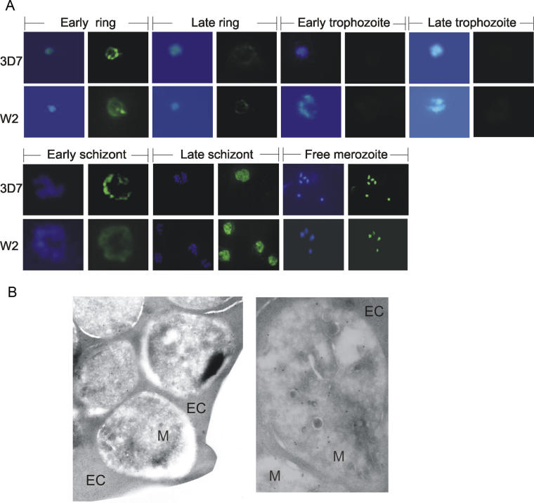
Erythrocytic malaria parasites utilize proteases for a number of cellular processes, including hydrolysis of hemoglobin, rupture of erythrocytes by mature schizonts, and subsequent invasion of erythrocytes by free merozoites. However, mechanisms used by malaria parasites to control protease activity have not been established. We report here the identification of an endogenous cysteine protease inhibitor of Plasmodium falciparum, falstatin, based on modest homology with the Trypanosoma cruzi cysteine protease inhibitor chagasin. Falstatin, expressed in Escherichia coli, was a potent reversible inhibitor of the P. falciparum cysteine proteases falcipain-2 and falcipain-3, as well as other parasite- and nonparasite-derived cysteine proteases, but it was a relatively weak inhibitor of the P. falciparum cysteine proteases falcipain-1 and dipeptidyl aminopeptidase 1. Falstatin is present in schizonts, merozoites, and rings, but not in trophozoites, the stage at which the cysteine protease activity of P. falciparum is maximal. Falstatin localizes to the periphery of rings and early schizonts, is diffusely expressed in late schizonts and merozoites, and is released upon the rupture of mature schizonts. Treatment of late schizionts with antibodies that blocked the inhibitory activity of falstatin against native and recombinant falcipain-2 and falcipain-3 dose-dependently decreased the subsequent invasion of erythrocytes by merozoites. These results suggest that P. falciparum requires expression of falstatin to limit proteolysis by certain host or parasite cysteine proteases during erythrocyte invasion. This mechanism of regulation of proteolysis suggests new strategies for the development of antimalarial agents that specifically disrupt erythrocyte invasion.
Conflict of interest statement
Figures


Similar articles
-
Cross-talk between malarial cysteine proteases and falstatin: the BC loop as a hot-spot target.PLoS One. 2014 Apr 3;9(4):e93008. doi: 10.1371/journal.pone.0093008. eCollection 2014. PLoS One. 2014. PMID: 24699522 Free PMC article.
-
Plasmodium falciparum cysteine protease falcipain-1 is not essential in erythrocytic stage malaria parasites.Proc Natl Acad Sci U S A. 2004 Jun 8;101(23):8721-6. doi: 10.1073/pnas.0402738101. Epub 2004 May 27. Proc Natl Acad Sci U S A. 2004. PMID: 15166288 Free PMC article.
-
Cysteine proteases of malaria parasites.Int J Parasitol. 2004 Dec;34(13-14):1489-99. doi: 10.1016/j.ijpara.2004.10.003. Int J Parasitol. 2004. PMID: 15582526 Review.
-
Falcipains and other cysteine proteases of malaria parasites.Adv Exp Med Biol. 2011;712:30-48. doi: 10.1007/978-1-4419-8414-2_3. Adv Exp Med Biol. 2011. PMID: 21660657 Review.
-
Deletion of the rodent malaria ortholog for falcipain-1 highlights differences between hepatic and blood stage merozoites.PLoS Pathog. 2017 Sep 18;13(9):e1006586. doi: 10.1371/journal.ppat.1006586. eCollection 2017 Sep. PLoS Pathog. 2017. PMID: 28922424 Free PMC article.
Cited by
-
The complex of Plasmodium falciparum falcipain-2 protease with an (E)-chalcone-based inhibitor highlights a novel, small, molecule-binding site.Malar J. 2019 Dec 2;18(1):388. doi: 10.1186/s12936-019-3043-0. Malar J. 2019. PMID: 31791339 Free PMC article.
-
Comparing sequence and structure of falcipains and human homologs at prodomain and catalytic active site for malarial peptide based inhibitor design.Malar J. 2019 May 3;18(1):159. doi: 10.1186/s12936-019-2790-2. Malar J. 2019. PMID: 31053072 Free PMC article.
-
The nucleotide specificity of succinyl-CoA synthetase of Plasmodium falciparum is not determined by charged gatekeeper residues alone.FEBS Open Bio. 2021 Mar;11(3):578-587. doi: 10.1002/2211-5463.13034. Epub 2021 Feb 17. FEBS Open Bio. 2021. PMID: 33174373 Free PMC article.
-
Identification of two new protective pre-erythrocytic malaria vaccine antigen candidates.Malar J. 2011 Mar 16;10:65. doi: 10.1186/1475-2875-10-65. Malar J. 2011. PMID: 21410955 Free PMC article.
-
Identification and characterization of the second cysteine protease inhibitor of Clonorchis sinensis (CsStefin-2).Parasitol Res. 2014 Jan;113(1):47-58. doi: 10.1007/s00436-013-3624-8. Epub 2013 Oct 8. Parasitol Res. 2014. PMID: 24100605
References
-
- Shenai BR, Sijwali PS, Singh A, Rosenthal PJ. Characterization of native and recombinant falcipain-2, a principal trophozoite cysteine protease and essential hemoglobinase of Plasmodium falciparum. J Biol Chem. 2000;275:29000–29010. - PubMed
-
- Singh N, Sijwali PS, Pandey KC, Rosenthal PJ. Plasmodium falciparum: Biochemical characterization of the cysteine protease falcipain-2′. Exp Parasitol. 2006;112:187–192. - PubMed
-
- Rosenthal PJ. Cysteine proteases of malaria parasites. Int J Parasitol. 2004;34:1489–1499. - PubMed
Publication types
MeSH terms
Substances
Associated data
- Actions
Grants and funding
LinkOut - more resources
Full Text Sources
Other Literature Sources
Molecular Biology Databases
