

Evaluation of S100A10, annexin II and B-FABP expression as markers for renal cell carcinoma
- PMID: 17083565
- PMCID: PMC11159138
- DOI: 10.1111/j.1349-7006.2006.00355.x
Evaluation of S100A10, annexin II and B-FABP expression as markers for renal cell carcinoma
Abstract
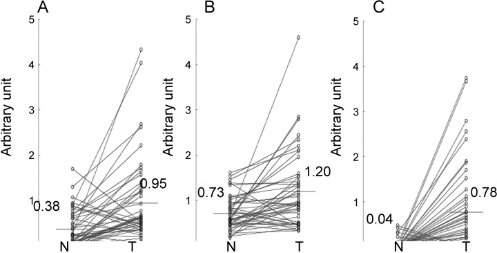
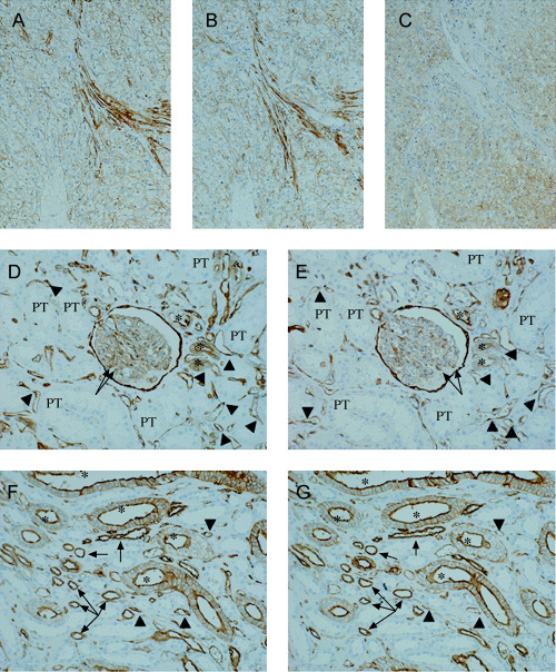
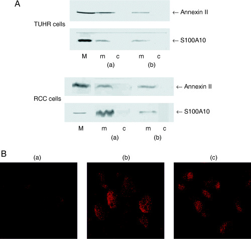
This study aimed to analyze expression of S100A10, annexin II and B-FABP genes in renal cell carcinoma (RCC) and their potential value as tumor markers. Furthermore, any correlation between the gene expression and prognostic indicators of RCC was analyzed. Expression of each gene was estimated by RT-PCR in the non-neoplastic (normal) and tumorous parts of resected kidney samples. Also, each antigen was immunostained in RCC and normal kidney tissues. Expression of the S100A10 gene averaged 2.5-fold higher in the tumor than that in the normal tissues (n = 47), after standardization against that of beta-actin. However, expression of annexin II, a natural ligand of S100A10, was only 1.64-fold higher. In the tissue sections of RCC, S100A10 and annexin II were immunostained in membranes. In the normal renal epithelia, however, both antigens were stained in the Bowman's capsule and the tubules from Henle's loop through the collecting duct system, but not in the proximal tubules, from where most RCC are derived. In contrast, expression of the B-FABP gene was 20-fold higher in the tumor. No B-FABP was immunohistochemically detected in normal kidney sections, but it was stained in the cytoplasm of RCC tissue sections. S100A10 and B-FABP genes were overexpressed regardless of nuclear grade and stage of RCC. Immunopositivity in RCC tissues (n = 13) was 100% for S100A10 and annexin II, and 70% for B-FABP; however, no clear relationship was observed in either antigen with nuclear grade and stage. It was found that all three performed well as RCC markers. B-FABP was most specific to RCC, as it was expressed little in normal kidney tissues.
Figures
References
-
- Teratani T, Watanabe T, Kuwahara F et al. Induced transcriptional expression of calcium‐binding protein S100A1 and S100A10 genes in human renal cell carcinoma. Cancer Lett 2002; 175: 71–7. - PubMed
-
- Teratani T, Watanabe T, Yamahara K et al. Restricted expression of calcium‐binding protein S100A5 in human kidney. Biochem Biophys Res Commun 2002; 291: 623–7. - PubMed
-
- Donato R. Functional roles of S100 proteins, calcium‐binding proteins of the EF‐hand type. Biochim Biophys Acta 1999; 1450: 191–231. - PubMed
-
- Heizmann CW, Fritz G, Schäfer W. S100 proteins: structure, functions and pathology. Frontiers Biosci 2002; 7: 1356–68. - PubMed
-
- Choi K‐S, Fogg DK, Yoon C‐S et al. p11 regulates extracellular plasmin production and invasiveness of HT1080 fibrosarcoma cells. FASEB J 2003; 17: 235–46. - PubMed
Publication types
MeSH terms
Substances
LinkOut - more resources
Full Text Sources
Other Literature Sources
Medical
Research Materials
