. 2006 Nov 3:3:78.
doi: 10.1186/1742-4690-3-78.

Phosphorylation of HIV-1 Tat by CDK2 in HIV-1 transcription

Affiliations

Phosphorylation of HIV-1 Tat by CDK2 in HIV-1 transcription

Tatyana Ammosova et al. Retrovirology. .

Abstract

Background: Transcription of HIV-1 genes is activated by HIV-1 Tat protein, which induces phosphorylation of RNA polymerase II (RNAPII) C-terminal domain (CTD) by CDK9/cyclin T1. Earlier we showed that CDK2/cyclin E phosphorylates HIV-1 Tat in vitro. We also showed that CDK2 induces HIV-1 transcription in vitro and that inhibition of CDK2 expression by RNA interference inhibits HIV-1 transcription and viral replication in cultured cells. In the present study, we analyzed whether Tat is phosphorylated in cultured cells by CDK2 and whether Tat phosphorylation has a regulatory effect on HIV-1 transcription.

Results: We analyzed HIV-1 Tat phosphorylation by CDK2 in vitro and identified Ser16 and Ser46 residues of Tat as potential phosphorylation sites. Tat was phosphorylated in HeLa cells infected with Tat-expressing adenovirus and metabolically labeled with 32P. CDK2-specific siRNA reduced the amount and the activity of cellular CDK2 and significantly decreased phosphorylation of Tat. Tat co-migrated with CDK2 on glycerol gradient and co-immunoprecipitated with CDK2 from the cellular extracts. Tat was phosphorylated on serine residues in vivo, and mutations of Ser16 and Ser46 residues of Tat reduced Tat phosphorylation in vivo. Mutation of Ser16 and Ser46 residues of Tat reduced HIV-1 transcription in transiently transfected cells. The mutations of Tat also inhibited HIV-1 viral replication and Tat phosphorylation in the context of the integrated HIV-1 provirus. Analysis of physiological importance of the S16QP(K/R)19 and S46YGR49 sequences of Tat showed that Ser16 and Ser46 and R49 residues are highly conserved whereas mutation of the (K/R)19 residue correlated with non-progression of HIV-1 disease.

Conclusion: Our results indicate for the first time that Tat is phosphorylated in vivo; Tat phosphorylation is likely to be mediated by CDK2; and phosphorylation of Tat is important for HIV-1 transcription.


Figures
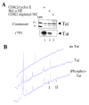
Figure 1
Figure 1
Analysis of HIV-1 Tat phosphorylated by CDK2/cyclin E in vitro. A, Tat is phosphorylated by CDK2. Recombinant Tat was phosphorylated in vitro by purified CDK2/cyclin E (lane 1), by HeLa nuclear extract (lane 2) or by CDK2-depleted HeLa nuclear extract (lane 3). Tat was resolved by 12% SDS Tris-Tricin PAGE. The gel was stained with Coomassie blue (upper panel) and exposed to Phospho Imager screen (lower panel). B, HPLC profiles of Tat peptides after trypsin cleavage. Recombinant Tat was phosphorylated in vitro by purified CDK2/cyclin E, resolved by 12% SDS Tris-Tricin PAGE, and subjected to in-gel trypsin digestion. The eluted peptides were resolved by reverse phase chromatography on μRPC C2/C18 ST 4.6/100 column. No Tat, mock trypsin digest without Tat. Tat, digest of non-phosphorylated Tat. (Phospho)-Tat, digestion of phosphorylated Tat. I and II, peaks identified in the elution profile of phosphorylated Tat that were subjected to MALDI TOF/TOF mass spectrometry.
Figure 2
Figure 2
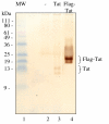
Expression of untagged and Flag-tagged Tat. COS-7 cells were transfected with Tat (lane 3) and Flag-tagged Tat (lane 4) expression vectors or mock-transfected (lane 2). At 48 hours post transfection cells were lysed and Tat was immediately immunoprecipitated with anti-Tat rabbit polyclonal antibodies (lanes 2–4). Immunoprecipitated Tat was resolved by 15% Tris-Tricine SDS-PAGE, transferred to polyvinylidene fluoride membrane and immunoblotted with anti-Tat monoclonal antibodies using the 3,3'-diaminobenzidine enhancer system. Positions of Tat and Flag-Tat are indicated. Lane 1, prestained 10 kDa molecular weight markers.
Figure 3
Figure 3
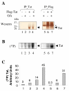
HIV-1 Tat is phosphorylated in cultured cells. HeLa cells were infected with recombinant adenovirus expressing Flag-tagged Tat as described in Methods (lanes 3, 4, 6 and 7). Lanes 1, 2, and 5 – control uninfected cells. At 48 hours post infection cells were labeled with (32P)-orthophosphate for 2 hours without (lanes 1, 3 and 6) or with (lanes 2, 4, 5 and 7) 1 μM okadaic acid (OA). Whole cell extracts were prepared and Tat was immunoprecipitated with anti-Tat rabbit polyclonal antibodies (lanes 1–4) or anti-Flag monoclonal murine antibodies (lanes 5–7). Immunoprecipitated Tat was resolved by 15% Tris-Tricine SDS-PAGE, and transferred to polyvinylidene fluoride membrane. A, immunoblot of the membrane with anti-Tat monoclonal antibodies using the 3,3'-diaminobenzidine enhancer system. B, autoradiography of the membrane on Phosphor Imager. C, quantification of panel B. The position of light chain of IgG recognized in anti-Flag immunoprecipitates by anti-mouse HRP-conjugated secondary antibodies is indicated by asterisk.
Figure 4
Figure 4
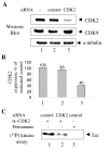
CDK2-directed siRNA inhibits CDK2 expression. A, CDK2-directed siRNA inhibits expression of CDK2. HeLa cells were transfected with siRNAs targeting CDK2 (lane 3) or non-targeting control pool (control, lane 2). Lane 1, untransfected cells. At 48 hours post-transfection cells were lysed and cellular extracts were resolved on 12% Tris-Tricine SDS-PAGE and analyzed by immunoblotting analysis with antibodies against CDK2, CDK9 or α-tubulin. B, quantification of the CDK2 expression in panel A using α-tubulin expression level for normalization. C, CDK2-directed siRNA inhibits enzymatic activity of CDK2. CDK2 was precipitated from cellular extracts prepared from HeLa cells transfected with siRNAs targeting CDK2 (lane 2) or non-targeting control (lanes 1 and 3). Lanes 1 and 2, precipitation with rabbit anti-CDK2 antibodies. Lane 3, precipitation with rabbit preimmune serum. Immunoprecipitates were incubated with γ-(32P)ATP and recombinant Tat (see Methods), resolved on 12% Tris-Tricine SDS-PAGE and analyzed by autoradiography on Phosphor Imager. Position of Tat is indicated.
Figure 5
Figure 5
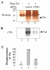
CDK2-directed siRNA blocks Tat phosphorylation. A, HeLa cells were infected with Adeno-Tat (lanes 2 and 3). At 4 hours post infection, cells were transfected with siRNAs targeting CDK2 (lane 3) or non-targeting control pool (lane 2). Lane 1 – control cells. At 48 hours post-infection cells were labeled with (32P)-orthophosphate for 2 hours. Whole cell extract was subjected to immunoprecipitation with anti-Flag antibodies, resolved by 15% Tris-Tricine SDS-PAGE, and transferred to polyvinylidene fluoride membrane. A, immunoblot of the membrane with anti-Tat monoclonal antibodies using the 3,3'-diaminobenzidine enhancer system. B, autoradiography of the membrane on Phosphor Imager screen. C, quantification of the panel B. Position of Tat is indicated by arrow. The position of light chain of IgG recognized in anti-Flag immunoprecipitates by anti-mouse HRP-conjugated secondary antibodies is indicated by asterisk.
Figure 6
Figure 6
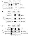
Tat and CDK2 co-migrate on glycerol gradient. 293T cell lysated from the cells infected with Adeno-Tat were fractionated on 10%–30% glycerol gradients by centrifugation and analyzed with indicated antibodies by Immunoblotting.
Figure 7
Figure 7
A, CDK2 associates with Tat in cultured cells. Flag-tagged Tat was expressed in HeLa cells by Adeno-Tat infection and precipitated with anti-Flag antibodies from cellular lysate. Co-precipitated proteins were resolved on 12% Tris-Tricine PAGE and analyzed by immunoblotting analysis with anti-CDK2 and anti-Tat antibodies. Lane 1, input control without Tat. Lane 2, input control with Tat. Lanes 3 and 4, extract without or with Tat precipitated with anti-Flag antibodies. Lane 5, extract with Tat precipitated with preimmune mouse IgG. The position of light chain of IgG recognized in anti-Flag immunoprecipitates by anti-mouse HRP-conjugated secondary antibodies is indicated by asterisk. B, CDK2-specific siRNA inhibits association of Tat with CDK2. Flag-tagged Tat was expressed in 293T cells by transfection (lanes 2 and 4). Cells were transfected with non-targeting (lanes 1 and 2) or CDK2-specific (lanes 3 and 4) siRNAs. Lysates were precipitated with anti-Flag antibodies, resolved on 12% Tris-Tricine PAGE and immunoblotted with anti-Tat, anti-CDK9 or anti-CDK2 antibodies. C, CDK2-specific siRNA does not affect association of Tat with cyclin T1. Flag-tagged Tat was expressed in 293T cells by transfection (lanes 3 and 5). Cells were transfected with non-targeting (lanes 2 and 3) or CDK2-specific (lanes 4 and 5) siRNAs. Lysates were precipitated with anti-Flag antibodies, resolved on 12% Tris-Tricine PAGE and immunoblotted with anti-Tat, anti-CDK9 or anti-CDK2 antibodies. Lane 1, input.
Figure 8
Figure 8
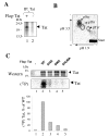
HIV-1 Tat is phosphorylated on S16 and S46 residues in vivo. A, HeLa cells were infected with recombinant adenovirus expressing Flag-tagged Tat as described in Methods. At 48 hours post infection cells were labeled with (32P)-orthophosphate for 2 hours with 1 μM okadaic acid (OA). Lane1, Flag-Tat was immunoprecipitated from whole cell extracts with anti-Flag antibodies and resolved by 15% Tris-Tricine SDS-PAGE. Lane 2, control mock-transfected cells. The picture is an autoradiogram. B, Tat peptides were eluted from the gel shown in panel A by overnight incubation with trypsin and subjected to acid hydrolysis as described in Materials and Methods. The hydrolyzed material was spotted on nitrocellulose plate and examined by two-dimensional thin layer electrophoresis and autoradiography. The indicated positions of non-radioactive phospho-amino acid standards were visualized by staining with 0.5% ninhydrin in ethanol. C, Mutations of S16 and S46 reduce Tat phosphorylation in vivo. 293T cells were transfected with vectors expressing Flag-tagged WT Tat (lane 2), Tat S16A (lane 3), Tat S46A (lane 4) or Tat S16,46A (lane 5). Lane 1, mock transfection. At 48 hours post-transfection the cells were labeled with (32P)-orthophosphate for 2 hours. Whole cell extract was subjected for immunoprecipitation with anti-Flag antibodies, resolved by 15% Tris-Tricine SDS-PAGE, and analyzed by Western blotting with polyclonal anti-Tat antibodies and on Phosphor Imager. Quantification is shown in the lower panel. Position of Tat is indicated by arrow.
Figure 9
Figure 9
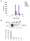
Mutations of Ser16 and Ser46 of Tat reduce its transactivation potential. A, COS-7 cells were transfected with WT Tat, Tat S16A, Tat S46A or Tat S16,46A expression vectors. At 48 hours posttransfection, the cells were lysed. Tat was immunoprecipitated from the lysates with rabbit polyclonal antibodies, resolved by 15% Tris-Tricine SDS-PAGE and immunoblotted with monoclonal anti-Tat antibodies. B, HeLa cells were transfected with the HIV-1 LTR-LacZ expression vector alone (not shown here) and in combination with WT Tat, Tat S16A, Tat S46A or Tat S16,46A expression vectors. At 48 hours posttransfection, cells were lysed and analyzed for β-galactosidase activity with ONPG.
Figure 10
Figure 10
A, Mutations of S16 and S46 of Tat reduce its ability to induce viral production. HLM1 is a HeLa derived cells containing one copy of integrated HIV-1 proviral genome with a Tat-defective mutation. Various Flag-tagged Tat (WT Tat, Tat S16A, Tat S46A or Tat S16,46A) expression vectors were used for HLM-1 transfections. Cells were cultured in complete media in absence of G418 for 14 days. The supernatants were collected at Day 0, 1, 2, 7 and 14, and analyzed for p24 by ELISA assay. B, Mutations of S16 and S46 of Tat inhibit Tat phosphorylation during one round of viral replication. HLM1 cells were transfected with vectors expressing Flag-tagged Tat (WT Tat, Tat S16A, Tat S46A or Tat S16,46A). At 48 hours post transfection the cells were labeled with (32P)-orthophosphate for 2 hours with 1 μM okadaic acid. Flag-Tat was immunoprecipitated from whole cell extracts with anti-Flag antibodies and resolved by 15% Tris-Tricine SDS-PAGE. The gel was dried and exposed to Phosphor Imager screen. Lane 1, Wt Tat. Lane 2, Tat S16A. Lane 3, Tat S46A. Lane 4, Tat S16,46A. Lane 5, mock-transfected cells. The picture is an autoradiogram.
Figure 11
Figure 11
Serine residues at position 16 and 46 are highly conserved in Tat isolates. The 158 sequences of Tat isolates deposited to PubMed database were analyzed for the presence of serines at position 16, 23, 46, 61, 68, 70, 73, 74 and 75. Presence of a serine at the indicated position is presented as a percent of the total number of the isolates analyzed.

References

    1. Brady J, Kashanchi F. Tat gets the "green" light on transcription initiation. Retrovirology. 2005;2:69. doi: 10.1186/1742-4690-2-69. - DOI - PMC - PubMed
    1. Brigati C, Giacca M, Noonan DM, Albini A. HIV Tat, its TARgets and the control of viral gene expression. FEMS Microbiol Lett. 2003;220:57–65. doi: 10.1016/S0378-1097(03)00067-3. - DOI - PubMed
    1. Coadou G, Gharbi-Benarous J, Megy S, Bertho G, Evrard-Todeschi N, Segeral E, Benarous R, Girault JP. NMR studies of the phosphorylation motif of the HIV-1 protein Vpu bound to the F-box protein beta-TrCP. Biochemistry. 2003;42:14741–14751. doi: 10.1021/bi035207u. - DOI - PubMed
    1. Agostini I, Popov S, Hao T, Li JH, Dubrovsky L, Chaika O, Chaika N, Lewis R, Bukrinsky M. Phosphorylation of Vpr regulates HIV type 1 nuclear import and macrophage infection. AIDS Res Hum Retroviruses. 2002;18:283–288. doi: 10.1089/088922202753472856. - DOI - PubMed
    1. Yang X, Gabuzda D. Mitogen-activated protein kinase phosphorylates and regulates the HIV-1 Vif protein. J Biol Chem. 1998;273:29879–29887. doi: 10.1074/jbc.273.45.29879. - DOI - PubMed

Publication types

LinkOut - more resources