Comparative Study
. 2006 Nov 3:7:74.
doi: 10.1186/1471-2202-7-74.

Estrogen protects neuronal cells from amyloid beta-induced apoptosis via regulation of mitochondrial proteins and function

Affiliations
Comparative Study

Estrogen protects neuronal cells from amyloid beta-induced apoptosis via regulation of mitochondrial proteins and function

Jon Nilsen et al. BMC Neurosci. .

Abstract

Background: Neurodegeneration in Alzheimer's disease is associated with increased apoptosis and parallels increased levels of amyloid beta, which can induce neuronal apoptosis. Estrogen exposure prior to neurotoxic insult of hippocampal neurons promotes neuronal defence and survival against neurodegenerative insults including amyloid beta. Although all underlying molecular mechanisms of amyloid beta neurotoxicity remain undetermined, mitochondrial dysfunction, including altered calcium homeostasis and Bcl-2 expression, are involved in neurodegenerative vulnerability.

Results: In this study, we investigated the mechanism of 17beta-estradiol-induced prevention of amyloid beta-induced apoptosis of rat hippocampal neuronal cultures. Estradiol treatment prior to amyloid beta exposure significantly reduced the number of apoptotic neurons and the associated rise in resting intracellular calcium levels. Amyloid beta exposure provoked down regulation of a key antiapoptotic protein, Bcl-2, and resulted in mitochondrial translocation of Bax, a protein known to promote cell death, and subsequent release of cytochrome c. E2 pretreatment inhibited the amyloid beta-induced decrease in Bcl-2 expression, translocation of Bax to the mitochondria and subsequent release of cytochrome c. Further implicating the mitochondria as a target of estradiol action, in vivo estradiol treatment enhanced the respiratory function of whole brain mitochondria. In addition, estradiol pretreatment protected isolated mitochondria against calcium-induced loss of respiratory function.

Conclusion: Therefore, we propose that estradiol pretreatment protects against amyloid beta neurotoxicity by limiting mitochondrial dysfunction via activation of antiapoptotic mechanisms.


Figures
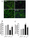
Figure 1
Figure 1
17β-estradiol prevents Amyloid beta1–42-induced neurotoxicity. Primary hippocampal neurons pretreated with E2 (10 ng/mL; 48 hr) or vehicle control were exposed to Aβ1–42 for 3 days and assessed for neuronal survival by calcein/ethidium homodimer staining. A, Visualization of viable (green) and dead (red) neurons. Bar = 20 μm. B-C, Fluorometer readings of calcein (live) and ethidium homodimer (dead) signals. (** = p < 0.01 as compared to control; ++ = p < 0.01 as compared to Aβ1–42 alone; n = 4).
Figure 2
Figure 2
17β-estradiol prevents Amyloid beta1–42-induced apoptosis. Primary hippocampal neurons pretreated with E2 (10 ng/mL; 48 hr) or vehicle control were exposed to Aβ1–42 for 3 days and assessed for apoptosis by TUNEL staining. A, Representative images of TUNEL positive neurons and total nuclei. Bar = 20 μm. B, Counts of TUNEL positive neurons normalized to total nuclei from 3 random fields per well are presented as mean ± SEM. (* = p < 0.05 as compared to control; + = p < 0.05 as compared to Aβ1–42 alone; n = 4).
Figure 3
Figure 3
17β-estradiol prevents Amyloid beta1–42-induced rise in resting calcium concentration. Primary hippocampal neurons pretreated with E2 (10 ng/mL; 48 hr) or vehicle control were exposed to Aβ1–42 for 24 hr and assessed for cytosolic calcium concentration by Fura2 imaging. A. Representative images of Fura2 fluorescence in psuedocolor representing 340/380 ratio. B. Quantitative changes in Fura2 340/380 ratio presented as means +/- S.E.M. (** = p < 0.01 as compared to control; ++ = p < 0.01 as compared to Aβ1–42 alone; n = 4).
Figure 4
Figure 4
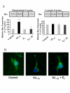
17β-estradiol prevents Amyloid beta1–42-induced decline in mitochondrial Bcl-2 expression and Bax translocation. Primary hippocampal neurons pretreated with E2 (10 ng/mL; 48 hr) or vehicle control were exposed to Aβ1–42 for 24 hr and mitochondrial fractions were assessed for (A) Bcl-2 and (B) Bax expression by Western blot analysis. (* = p < 0.05 as compared to control; ** = p < 0.01 as compared to control; + = p < 0.05 as compared to Aβ1–42 alone; n = 4).
Figure 5
Figure 5
17β-estradiol prevents Amyloid beta1–42-induced Bax translocation. Primary hippocampal neurons pretreated with E2 (10 ng/mL; 48 hr) or vehicle control were exposed to Aβ1–42 for 24 hr and mitochondrial fractions were assessed for Bax expression by (A) Western blot analysis and (B) immunocytochemistry. The order of bands was switched to maintain a consistent treatment order across all figures for presentation purposes only. All quantitation and analysis was performed unmodified images. (* = p < 0.05 as compared to control; ** = p < 0.01 as compared to control; + = p < 0.05 as compared to Aβ1–42 alone; n = 4).
Figure 6
Figure 6
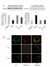
17β-estradiol prevents Amyloid beta1–42-induced cytochrome c release. Primary hippocampal neurons pretreated with E2 (10 ng/mL; 48 hr) or vehicle control were exposed to Aβ1–42 for 24 hr and assessed for cytochrome c expression by Western blot analysis and immunocytochemistry. A, Western blot analysis cytochrome c expression in cytosolic (right panel) and mitochondrial (left panel) fractions of treated neurons. B, Treated neurons were immunostained for cytochrome c expression (Green) and mitochondria were labeled with Mitotracker Red CMXRos (Red). Co-localization of cytochrome c with mitochondria is visible in the overlay image (yellow). Bar = 5 μm.
Figure 7
Figure 7
17β-estradiol prevents Amyloid beta1–42-induced Bax translocation and cytochrome c release. Primary hippocampal neurons pretreated with E2 (10 ng/mL; 48 hr) or vehicle control were exposed to Aβ1–42 for 24 hr and assessed for Bax and cytochrome c expression by immunocytochemistry. Treated neurons were double immunostained for Bax (Red) and cytochrome c expression (Green). Bar = 10 μm.
Figure 8
Figure 8
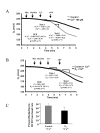
17β-estradiol prevents calcium-induced damage to mitochondrial respiratory function. A. Whole brain mitochondria were isolated from ovariectomized rats and assessed for mitochondrial respiration in the presence of glutamate (5 μM) and malate (5 μM) as respiratory substrates following a 2 min challenge with 100 μM Ca2+ or buffer control. Respiratory control ratio (RCR) is the ratio of State 3 (ADP stimulated) respiration to State 4 (resting) respiration. (n = 7; * = p < 0.05) B. Whole brain mitochondria were isolated from ovariectomized rats treated with 17β-estradiol (30 μg/kg) or vehicle control for 24 hr and assessed for mitochondrial respiration following 2 min calcium (10 μM) exposure in the presence of glutamate (5 μM) and malate (5 μM) as respiratory substrates. C. Respiratory control ratio (RCR) is the ratio of State 3 (ADP stimulated) respiration to State 4 (resting) respiration. (n = 7; * = p < 0.05)

References

    1. Tan S, Wood M, Maher P. Oxidative stress induces a form of programmed cell death with characteristics of both apoptosis and necrosis in neuronal cells. Journal of Neurochemistry. 1998;71:95–105. - PubMed
    1. LaFerla FM, Tinkle BT, Bieberich CJ, Haudenschild CC, Jay G. The Alzheimer's A beta peptide induces neurodegeneration and apoptotic cell death in transgenic mice. Nat Genet. 1995;9:21–30. doi: 10.1038/ng0195-21. - DOI - PubMed
    1. Shimohama S. Apoptosis in Alzheimer's disease--an update. Apoptosis. 2000;5:9–16. doi: 10.1023/A:1009625323388. - DOI - PubMed
    1. LaFerla FM. Calcium dyshomeostasis and intracellular signalling in Alzheimer's disease. Nat Rev Neurosci. 2002;3:862–872. doi: 10.1038/nrn960. - DOI - PubMed
    1. Landfield PW, Thibault O, Mazzanti ML, Porter NM, Kerr DS. Mechanisms of neuronal death in brain aging and Alzheimer's disease: role of endocrine-mediated calcium dyshomeostasis. J Neurobiol. 1992;23:1247–1260. doi: 10.1002/neu.480230914. - DOI - PubMed

Publication types

MeSH terms