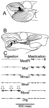
Masticatory muscles and the skull: a comparative perspective
- PMID: 17084804
- PMCID: PMC1853336
- DOI: 10.1016/j.archoralbio.2006.09.010
Masticatory muscles and the skull: a comparative perspective
Abstract
Masticatory muscles are anatomically and functionally complex in all mammals, but relative sizes, orientation of action lines, and fascial subdivisions vary greatly among different species in association with their particular patterns of occlusion and jaw movement. The most common contraction pattern for moving the jaw laterally involves a force couple of protrusor muscles on one side and retrusors on the other. Such asymmetrical muscle usage sets up torques on the skull and combines with occlusal loads to produce bony deformations not only in the tooth-bearing jaw bones, but also in more distant elements such as the braincase. Maintenance of bone in the jaw joint, and probably elsewhere in the skull, is dependent on these loads.
Figures


Similar articles
-
Dry versus wet and gross: Comparisons between the dry skull method and gross dissection in estimations of jaw muscle cross-sectional area and bite forces in sea otters.J Morphol. 2019 Nov;280(11):1706-1713. doi: 10.1002/jmor.21061. Epub 2019 Sep 12. J Morphol. 2019. PMID: 31513299
-
Jaw muscles and the skull in mammals: the biomechanics of mastication.Comp Biochem Physiol A Mol Integr Physiol. 2001 Dec;131(1):207-19. doi: 10.1016/s1095-6433(01)00472-x. Comp Biochem Physiol A Mol Integr Physiol. 2001. PMID: 11733178 Review.
-
The relationship between skull morphology, masticatory muscle force and cranial skeletal deformation during biting.Ann Anat. 2016 Jan;203:59-68. doi: 10.1016/j.aanat.2015.03.002. Epub 2015 Mar 21. Ann Anat. 2016. PMID: 25829126
-
[Masticatory muscles. Part III. Biomechanics of the masticatory muscles].Ned Tijdschr Tandheelkd. 1997 Aug;104(8):302-5. Ned Tijdschr Tandheelkd. 1997. PMID: 11924415 Dutch.
-
Dynamics of the human masticatory system.Crit Rev Oral Biol Med. 2002;13(4):366-76. doi: 10.1177/154411130201300406. Crit Rev Oral Biol Med. 2002. PMID: 12191962 Review.
Cited by
-
Therapeutic effect of localized vibration on alveolar bone of osteoporotic rats.PLoS One. 2019 Jan 29;14(1):e0211004. doi: 10.1371/journal.pone.0211004. eCollection 2019. PLoS One. 2019. PMID: 30695073 Free PMC article.
-
Heterochrony and patterns of cranial suture closure in hystricognath rodents.J Anat. 2009 Mar;214(3):339-54. doi: 10.1111/j.1469-7580.2008.01031.x. J Anat. 2009. PMID: 19245501 Free PMC article.
-
Engineering a targeted and safe bone anabolic gene therapy to treat osteoporosis in alveolar bone loss.Mol Ther. 2024 Sep 4;32(9):3080-3100. doi: 10.1016/j.ymthe.2024.06.036. Epub 2024 Jun 26. Mol Ther. 2024. PMID: 38937970 Free PMC article.
-
Adaptation in the Alleyways: Candidate Genes Under Potential Selection in Urban Coyotes.Genome Biol Evol. 2025 Jan 6;17(1):evae279. doi: 10.1093/gbe/evae279. Genome Biol Evol. 2025. PMID: 39786569 Free PMC article.
-
Osteogenic effect of high-frequency acceleration on alveolar bone.J Dent Res. 2012 Apr;91(4):413-9. doi: 10.1177/0022034512438590. Epub 2012 Feb 14. J Dent Res. 2012. PMID: 22337699 Free PMC article.
References
-
- Turnbull WD. Mammalian masticatory apparatus. Fieldiana: Geology. 1970;18:149–356.
-
- Herring SW, Grimm AF, Grimm BR. Functional heterogeneity in a multipinnate muscle. Am J Anat. 1979;154:563–576. - PubMed
-
- Wood WW. A review of masticatory muscle function. J Prosth Dent. 1987;57:222–232. - PubMed
-
- Blanksma NG, van Eijden TMGJ, van Ruijven LJ, Weijs WA. Electromyographic heterogeneity in the human temporalis and masseter muscles during dynamic tasks guided by visual feedback. J Dent Res. 1997;76:542–551. - PubMed
-
- Turkawski SJJ, Van Eijden TMGJ, Weijs WA. Force vectors of single motor units in a multipennate muscle. J Dent Res. 1998;77:1823–1831. - PubMed
Publication types
MeSH terms
Grants and funding
LinkOut - more resources
Full Text Sources

