. 2007 Feb 15;302(2):412-26.
doi: 10.1016/j.ydbio.2006.09.044. Epub 2006 Oct 3.

Regulation of the Drosophila distal antennal determinant spineless

Affiliations

Regulation of the Drosophila distal antennal determinant spineless

Richard B Emmons et al. Dev Biol. .

Abstract

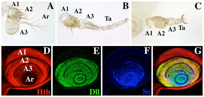
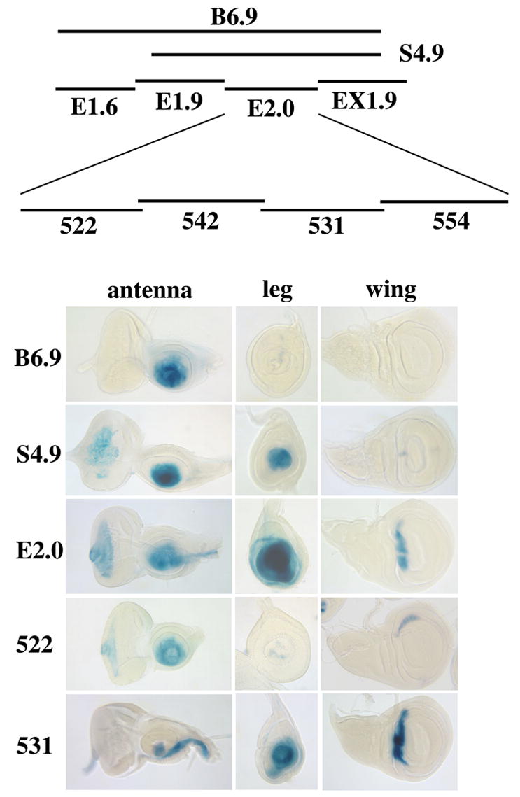
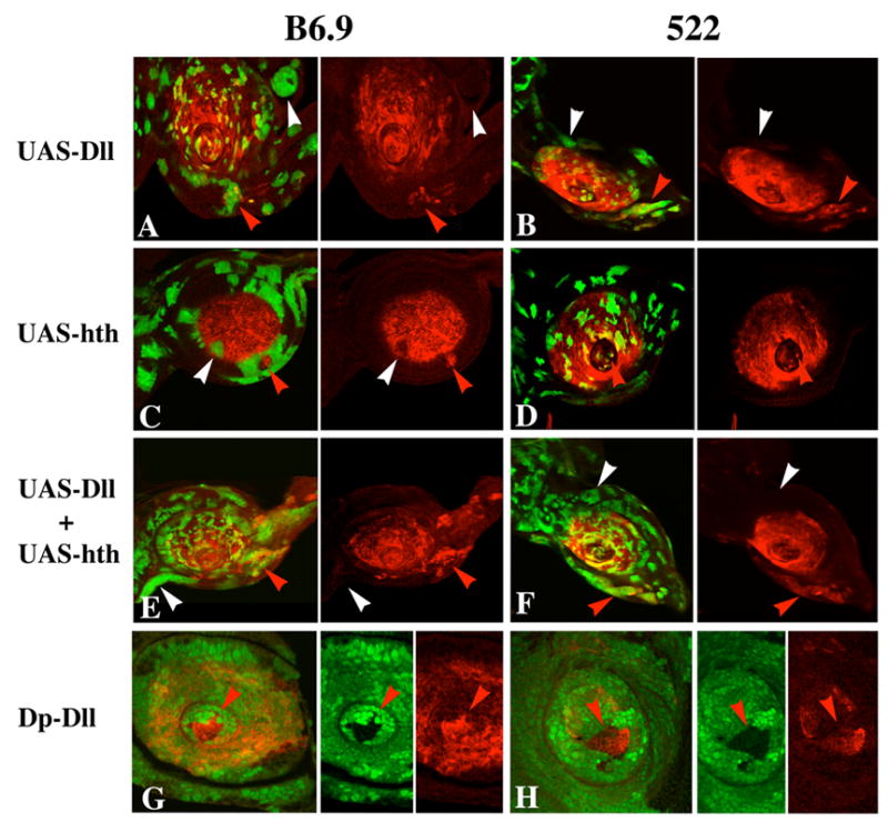
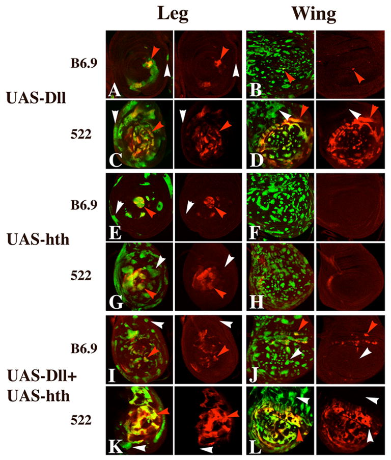
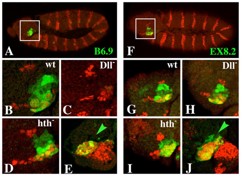
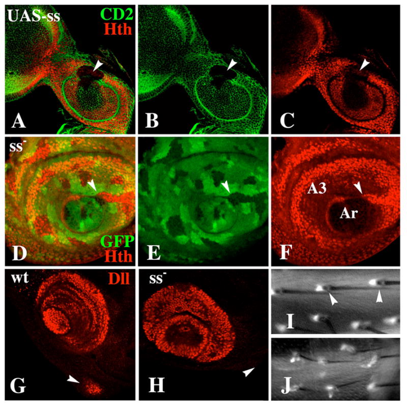
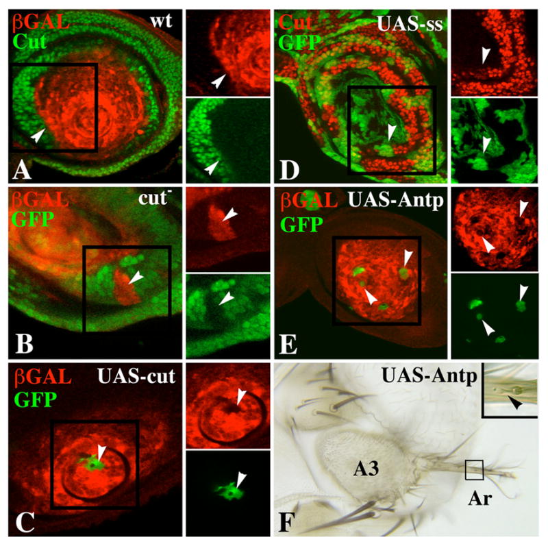
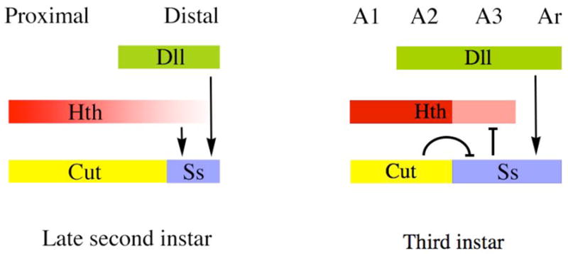
The transformation of antenna to leg is a classical model for understanding segmental fate decisions in Drosophila. The spineless (ss) gene encodes a bHLH-PAS transcription factor that plays a key role in specifying the identity of distal antennal segments. In this report, we identify the antennal disc enhancer of ss and then use enhancer-lacZ reporters to work out how ss antennal expression is regulated. The antennal determinants Distal-less (Dll) and homothorax (hth) are key activators of the antennal enhancer. Dll is required continuously and, when present at elevated levels, can activate the enhancer in regions devoid of hth expression. In contrast, homothorax (hth) is required only transiently both for activation of the enhancer and for specification of the aristal portion of the antenna. The antennal enhancer is repressed by cut, which determines its proximal limit of expression, and by ectopic Antennapedia (Antp). Repression by Antp is not mediated by hth, suggesting that ss may be a direct target of Antp. Finally, we show that ss+ is not a purely passive target of its regulators: ss+ partially represses hth in the third antennal segment and lies upstream of Dll in the development of the maxillary palp primordia.


Figures

Figure 1
Figure 1
Antennae from wild-type and ss mutant adults, and expression of Hth, Dll, and the ss antennal reporter B6.9 in a mature antennal disc. A) A wild-type antenna. The first (A1), second (A2), and third (A3) antennal segments and the arista (Ar) are indicated. B) Antenna from an ssa-type mutant (ssD114.7/Df(3R)ssD114.4). Note that distal A3 and the arista are transformed to an almost complete set of tarsi (Ta). C) Antenna from a ss null mutant (ssD115.7/Df(3R)ssD114.4). Apart from a reduction in bristle size, A1 and A2 are basically normal; A3 is composed of naked cuticle, and the arista is reduced to a fifth tarsal segment (Ta) with claws. Panels D–G show a late third instar antennal disc triply labeled for Hth (red), Dll (green), and the ss antennal reporter B6.9 (blue). The primordia of A1, A2, A3, and Ar are indicated. Note that Hth (red) is present at a lower level in A3 than in A1 and A2, and is absent in the aristal region (D), Dll (green) is expressed in A2 and more distally (E), and the B6.9 ss antennal reporter (blue) is expressed in A3 and more distally (F). G) Merging of panels D–F.
Figure 2
Figure 2
Enhancer mapping within ss. The DNA scale is marked in kilobases. Above this scale are shown the ss transcribed region (exons are indicated as blue boxes; coding regions are dark blue, noncoding regions are light blue) and the locations of four ss mutants (A designates antennal transformations and B designates bristle reduction). The fragments tested for enhancer activity are indicated in red, and the expression patterns each drives are shown below for embryos (top), imaginal discs (middle) and for structures at the pupal stage (bottom). Embryonic expression is shown for only B6.9 and EX8.2. The expression patterns shown for EX6.5 are actually those of its P732 subfragment, which drives persistent, rather than transient, expression in the leg and antenna (see results). For all fragments, the embryonic and disc expression patterns shown correspond to patterns detected for the ss transcript (Duncan et al. 1998). Abbreviations: Ab (abdominal tergite), An (antenna), E (embryo), L (leg), P (maxillary palp), W (wing).
Figure 3
Figure 3
Dissection of the B6.9 fragment. Top: Summary of fragments tested using lacZ reporters. Bottom: X-Gal stained eye-antennal, leg, and wing discs showing the expression patterns driven by each fragment. B6.9 drives expression in the distal antenna, but not in leg or wing discs. In contrast, the S4.9 and E2.0 subfragments drive expression strongly in both antennal and leg discs. E2.0 also drives expression in a ventral stripe in the antennal disc and in the dorsal hinge region of the wing disc. S4.9 shows very weak expression in this same region of the wing disc. The leg and antennal disc expression of E2.0 prove to be largely separable, with the 522 and 531 fragments driving expression primarily in the distal antenna and leg, respectively. The separation is not complete, however, with 522 driving weak mottled expression in the distal leg, and 531 driving expression in the aristal region and in a ventral stripe in the antennal disc. 522 and 531 also drive expression in wing discs in the lateral and dorsal hinge regions, respectively. The E1.6, E1.9, EX1.9, 542 and 554 fragments do not drive expression in antennal or leg discs.
Figure 4
Figure 4
Regulation of the B6.9 and 522 antennal reporters by Dll and hth. In all discs shown here, loss of GFP fluorescence (green) is used to mark clones, and β-galactosidase expression is shown in red. A, E: Dependence of B6.9 (A) and 522 (E) expression on Dll+. Expression of both reporters is lost within Dll clones (arrowheads). This is true for clones induced throughout larval development. B–H: Control of B6.9 (B, C, D) and 522 (F, G, H) expression by hth. The images in B, C, and D are complicated by the fact that the B6.9 insertion studied is located in the same chromosome arm (3R) as hth. Consequently, the hth+/hth+ twin spot clones (recognized by strong green fluorescence) lack the B6.9 reporter altogether. B, F: hth clones induced 1–2 days AEL. Such clones lose expression of B6.9 and 522 autonomously (arrowheads). C, G: hth clones induced 2–3 days AEL. Such clones show persistent expression of B6.9 in the aristal region (arrow), but loss of expression more proximally (arrowhead in C). 522 expression is usually lost in such clones, although some persistence is seen (arrows in G). D, H: hth clones induced 4–5 days AEL. B6.9 expression is unaffected (arrows in D), whereas 522 expression is lost in some clones (arrowhead in H) but retained in others (arrows in H). I–L: Effects on antennal identity of hth clones induced at different times of development. I: A normal antenna heterozygous for the bristle markers (M(3)w Bsb) used to mark hth clones. Note the reduction of the aristal (Ar) branches. J: An antenna containing hth clones induced during the second day AEL [time corrected for the delay caused by heterozygosity for M(3)w]. Because the Minute technique was used to confer a growth advantage to clones, the antenna is probably entirely mutant. The entire antenna is transformed to leg, with the distal region developing as a set of tarsal segments (Ta) terminated by claws (Cl). K: An antenna containing hth clones induced during the third day AEL. Note that partially formed tarsal segments are present, terminated by an arista (arrow). L: An antenna containing hth clones induced during the fourth day AEL. Note that tarsal segments are absent, and that a complete arista including a basal cylinder (arrow) is present.
Figure 5
Figure 5
Effect of ectopic expression of Dll and Hth on expression of B6.9 and 522 in antennal discs. In all panels, clones are marked by GFP fluorescence (green) and β-galactosidase expression is in red. A, B: Dll-expressing clones induce expression of both B6.9 (A) and 522 (B) in the region ventral to the antenna (red arrowheads), but not elsewhere (white arrowheads). C, D: Hth-expressing clones often repress B6.9 and 522 proximally (white arrowheads), but upregulate 522 when located in the most distal (aristal) region (red arrowhead in D). Note that proximal Hth-expressing clones tend to follow the proximal limit of reporter expression (the A2–A3 boundary) for many cells (C). The disc shown in C is unusual in that a patch of β-galactosidase expressing cells is present proximal to the normal expression domain of the reporter (red arrowhead); whether this results from new induction of expression by the surrounding cells or from entrapment of originally distal cells is not clear. E, F: Clones expressing both Dll and Hth are very similar to clones expressing only Dll in their effects on B6.9 and 522 expression. G, H: Effects of varying Dll dosage on expression of 522 in antennal (G) and leg (H) discs. These discs contain clones carrying four doses of Dll+ (no green fluorescence) and two doses of Dll+ (bright green fluorescence), in a background of cells carrying three doses (light green fluorescence). Note the strong activation of 522 in the four-dose clones in both the antenna and leg (red arrowheads).
Figure 6
Figure 6
Effect of ectopic expression of Dll and Hth on expression of B6.9 and 522 in leg and wing discs. A–D: Dll-expressing clones activate 522 expression strongly in the distal leg (C) and in the wing blade (D) (red arrowheads), but not in more proximal regions of either disc (white arrowheads). Such clones can also activate B6.9 in the distal leg (red arrowhead in A) and, rarely, in the wing (red arrowhead in B). E–H: Hth-expressing clones also frequently activate both reporters in the distal leg (red arrowheads in E and G), but do not activate the reporters in the proximal leg (white arrowheads in E and G) or anywhere in the wing blade (F and H). I–L: Clones expressing both Dll and Hth behave much like clones expressing only Dll, with the exception that B6.9 is weakly activated in the wing hinge region in clones expressing both proteins (J).
Figure 7
Figure 7
Expression of the B6.9 and EX8.2 ss antennal reporters in embryos. β-galactosidase is stained green in all panels. Engrailed (which marks the posterior of each body segment) is stained red in all panels except E and J, in which Dll is stained red. Embryos shown in panels E and J are at stage 13; all others are at stage 12. A–E: Expression of B6.9. A: B6.9 is expressed in the antennal segment (inset) and in a single lateral cell in each segment. B: A higher magnification of the inset region from another embryo. Antennal expression of B6.9 is lost in Dll embryos (C), but is unaffected in hth embryos (D). E: Expression of B6.9 in the antennal segment coincides precisely with that of Dll. F–J: Expression of EX8.2. Note that expression extends from one edge of the antennal segment to the other (F+G) (like expression of ss) and is unaffected in Dll (H) and hth (I) embryos. J: EX8.2 is expressed in all Dll-expressing cells in the antennal segment as well as a few more anterior cells (green arrow). B6.9 does not drive expression in these anterior cells (green arrow in E).
Figure 8
Figure 8
Effect of ss on expression of hth and Dll. A–C: A Ss-expressing clone (marked by loss of CD2) causes repression of hth in the antenna (arrowhead). D–F: A ss clone causes increased expression of hth in A3 (arrowhead). G+H: Expression of Dll in wild-type and ss antennal discs. In wild type, Dll expression in the maxillary palp primordium (arrowhead) is activated in the prepupa (G). In ss mutants, Dll fails to be expressed in the palp primordium at this time (H). I+J: Expression of Dll-GAL4/UAS-GFP in ss+ (I) and ss (J) adult femurs. (I) In wild type, Dll is expressed in a single cell associated with each bract (arrowheads). (J) In ss mutants, bracts are absent from the femur, and Dll is expressed weakly in elongate cells that are associated with most bristles.
Figure 9
Figure 9
Mutual repression of ss and cut, and repression of the ss antennal reporters by ectopic Antp. A. Antennal disc double-labeled for Cut (green) and B6.9 expression (red) showing that the two expression patterns abut at the A2–A3 boundary (white arrowheads). B. A cut clone (arrowhead) marked by loss of GFP fluorescence (green) shows ectopic expression of B6.9 (red). C. A clone expressing Cut ectopically (green) represses B6.9 expression (arrowheads). D. Clones expressing ss ectopically (green) partially repress cut expression in the proximal antenna (arrowheads). E. Clones expressing Antp ectopically (green) reduce B6.9 expression (arrowheads). These clones were induced 4–5 days AEL. F. Antenna containing Antp-expressing clone(s) induced 3–4 days AEL. The clone is marked by yellow. Note partial transformation of arista (Ar) to tarsus. Bristles on the partially transformed arista are bracted (arrowhead), indicating leg identity.
Figure 10
Figure 10
Summary of regulatory interactions among hth, Dll, cut, and ss. On the left are shown proximo-distal expression patterns in the late second instar, shortly after Dll and ss (B6.9) have become activated in the antennal disc and when hth expression is diminished distally. At this time, ss is activated by both hth and Dll. In the third instar (right), the ss antennal enhancer continues to be activated by Dll, but no longer requires hth for its expression. Instead, ss weakly represses hth, resulting in its down-regulation in A3. Repression by cut defines the proximal limit of ss expression during the third instar; regulatory interactions between cut and ss during the second instar have not been examined.

Similar articles

Cited by

References

    1. Abu-Shaar M, Mann RS. Generation of multiple antagonistic domains along the proximodistal axis during Drosophila leg development. Development. 1998;125:3821–3830. - PubMed
    1. Abzhanov A, Kaufman TC. Homologs of Drosophila appendage genes in the patterning of arthropod limbs. Dev Biol. 2000;227:673–689. - PubMed
    1. Adachi-Yamada T, Harumoto T, Sakurai K, Ueda R, Saigo K, O’Connor MB, Nakato H. Wing-to-leg homeosis by spineless causes apoptosis regulated by fish-lips, a novel leucine-rich repeat transmembrane protein. Molec Cell Biol. 2005;25:3140–3150. - PMC - PubMed
    1. Angelini DR, Kaufman TC. Functional analyses in the hemipteran Oncopeltus fasciatus reveal conserved and derived aspects of appendage patterning in insects. Dev Biol. 2004;271:306–321. - PubMed
    1. Burgess EA, Duncan I. Direct control of antennal identity by the spineless-aristapedia gene of Drosophila. Mol Gen Genet. 1990;221:347–352. - PubMed

Publication types

MeSH terms

LinkOut - more resources