Comparative Study
. 2007 Mar 30;360(1):105-19.
doi: 10.1016/j.virol.2006.09.036. Epub 2006 Nov 7.

Macrophage entry mediated by HIV Envs from brain and lymphoid tissues is determined by the capacity to use low CD4 levels and overall efficiency of fusion

Affiliations
Comparative Study

Macrophage entry mediated by HIV Envs from brain and lymphoid tissues is determined by the capacity to use low CD4 levels and overall efficiency of fusion

Elaine R Thomas et al. Virology. .

Abstract

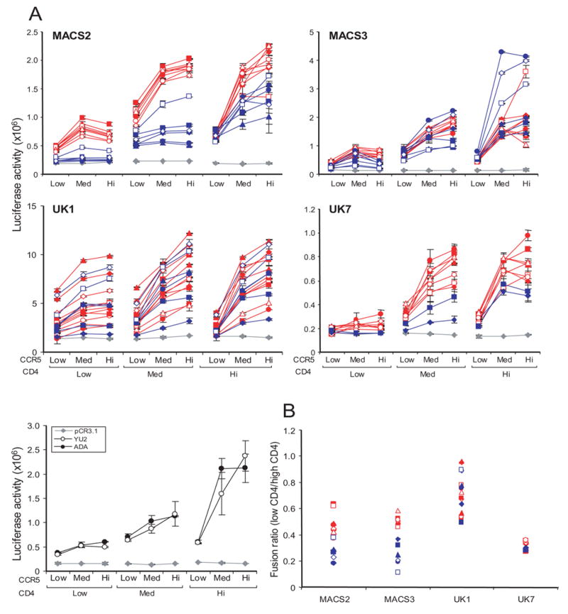
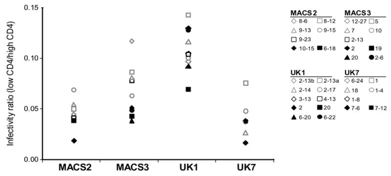
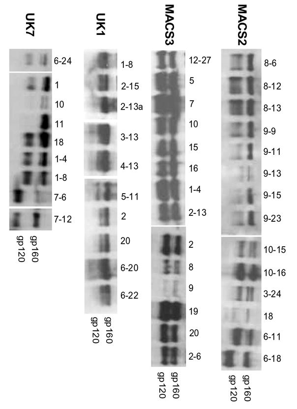
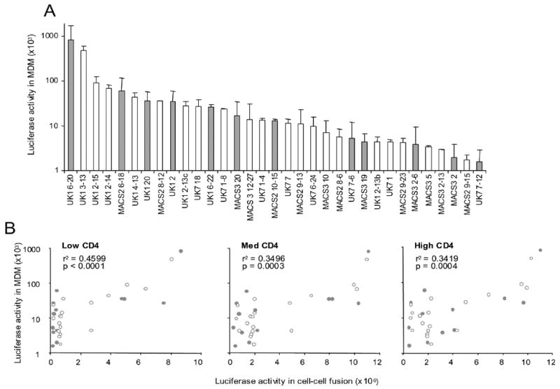
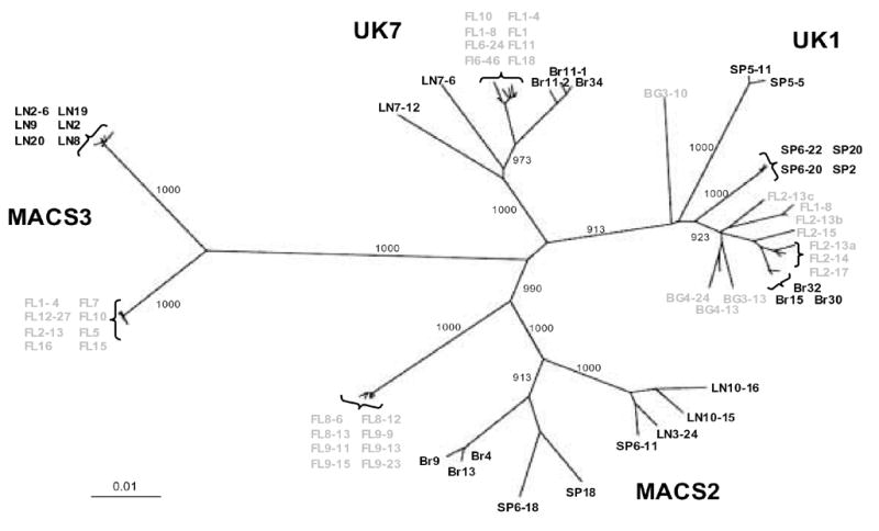
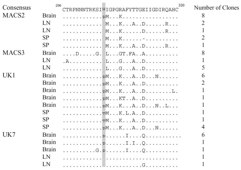
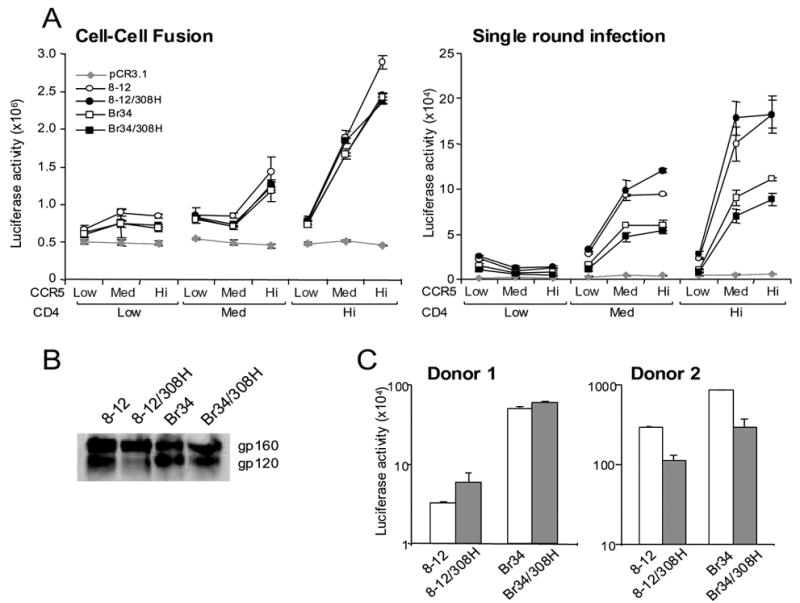
HIV infects macrophages and microglia in the central nervous system (CNS), which express lower levels of CD4 than CD4+ T cells in peripheral blood. To investigate mechanisms of HIV neurotropism, full-length env genes were cloned from autopsy brain and lymphoid tissues from 4 AIDS patients with HIV-associated dementia (HAD). Characterization of 55 functional Env clones demonstrated that Envs with reduced dependence on CD4 for fusion and viral entry are more frequent in brain compared to lymphoid tissue. Envs that mediated efficient entry into macrophages were frequent in brain but were also present in lymphoid tissue. For most Envs, entry into macrophages correlated with overall fusion activity at all levels of CD4 and CCR5. gp160 nucleotide sequences were compartmentalized in brain versus lymphoid tissue within each patient. Proline at position 308 in the V3 loop of gp120 was associated with brain compartmentalization in 3 patients, but mutagenesis studies suggested that P308 alone does not contribute to reduced CD4 dependence or macrophage-tropism. These results suggest that HIV adaptation to replicate in the CNS selects for Envs with reduced CD4 dependence and increased fusion activity. Macrophage-tropic Envs are frequent in brain but are also present in lymphoid tissues of AIDS patients with HAD, and entry into macrophages in the CNS and other tissues is dependent on the ability to use low receptor levels and overall efficiency of fusion.


Figures

Fig. 1
Fig. 1. Cell-cell fusion mediated by brain and lymphoid Envs
(A) 293T cells expressing brain (red) or lymphoid (blue) Envs from MACS2, MACS3, UK1, and UK7 or the ADA or YU2 Envs were mixed with Cf2luc cells expressing low, medium (Med), or high (Hi) levels of CD4 and CCR5 as indicated. Fusion was measured as luciferase counts in lysed cells following 12 h incubation. Control fusion of 293T cells with pCR3.1 in place of Env, indicating background levels, is represented by grey diamonds. Luciferase activity less than 1.5-fold above the background level obtained with the no Env negative control (pCR3.1) was considered negative. Data are expressed as means of duplicate wells from a single experiment and are representative of results from 2 to 3 independent experiments. Error bars represent standard deviations. (B) Fusion activity of brain (open symbols) and lymphoid (closed symbols) Envs from MACS2, MACS3, UK1 and UK7 expressed as a ratio of Env-mediated fusion with cells expressing low compared to high levels of CD4 and medium levels of CCR5.
Fig. 2
Fig. 2. Virus entry mediated by brain and lymphoid Envs
Luciferase reporter viruses were pseudotyped with brain (open symbols) or lymphoid (closed symbols) Envs from MACS2, MACS3, UK1 and UK7. Entry into Cf2 cells is expressed as a ratio of virus entry on cells expressing low compared to high levels of CD4 and high levels of CCR5. Actual values for luciferase counts in cells expressing low or high CD4 ranged from 823-101393, 788-34703, 1053-1072087, and 315-58126 for Envs from MACS2, MACS3, UK1 and UK7, respectively. Background levels were determined as in Fig. 1.
Fig. 3
Fig. 3. gp160 and gp120 expression by brain and lymphoid Env clones
Env expression and processing of gp160 to gp120 in 293T cells detected by Western blot with anti-gp120 rabbit sera. Positions of gp160 and gp120 bands are indicated on the right.
Fig. 4
Fig. 4. Virus entry into macrophages mediated by brain and lymphoid Envs
(A) Luciferase reporter viruses were pseudotyped with brain (open bars) or lymphoid (grey bars) Envs from MACS2, MACS3, UK1 and UK7. Entry into macrophages was measured as luciferase counts in lysed cells following incubation for 6 days. Data are expressed as means from duplicate wells and are representative of results in 2 different donors. Error bars represent standard deviations. (B) Brain (open symbols) and lymphoid (grey symbols) entry into MDM (y axis) correlates with levels of fusion between Env-expressing 293T cells and Cf2luc cells expressing low, medium, or high levels of CD4 and high CCR5 (x axis). Pearson's correlation coefficient (r2) and p values are shown.
Fig. 5
Fig. 5. Phylogenetic analysis of full-length HIV env nucleotide sequences from autopsy tissue samples and viral isolates
Env sequences amplified directly from brain and lymphoid tissues are color-coded grey and black, respectively. Env sequences for MACS2, UK1 and UK7 brain viral isolates obtained by PBMC co-culture are shown in black with the prefix Br. Numbers associated with each branch are bootstrap values, which represent the number of trees, out of 1000 replicates performed, in which the same branching order was found. Only values above 900 for the major branches are shown. Branch lengths are proportional to the amount of sequence divergence. Scale bars indicate 1% sequence divergence. FL, frontal lobe; BG, basal anglia; LN, lymph node; SP, spleen.
Fig. 6
Fig. 6. Amino acid alignment of HIV Env V3 loop regions of brain and lymphoid Env clones with the clade B consensus
Tissue of origin and number of clones with each sequence are indicated to the left and right of each sequence, respectively. Numbering is relative to the Hxbc2 Env sequence. Dots indicate residues identical to the clade B consensus, and dashes indicate gaps. Proline at position 308 is shaded in grey. LN, lymph node; SP, spleen.
Fig. 7
Fig. 7. Fusion and entry of brain Envs with a proline to histidine substitution at position 308
(A) 293T cells or luciferase reporter viruses pseudotyped with wild type and mutant brain Envs from MACS2 (8–12) and UK7 (Br34) were mixed with Cf2luc or Cf2 cells, respectively, expressing low, medium, or high levels of CD4 and CCR5 as indicated. Fusion and infection were measured as luciferase counts in lysed cells following 12 or 72 h incubation, respectively. Background levels were determined as in Fig. 1. (B) Wild type and mutant Env expression and processing of gp160 to gp120 in 293T cells detected by Western blot with anti-gp120 rabbit sera. Positions of gp160 and gp120 bands are indicated on the right. (C) Luciferase reporter viruses pseudotyped with wild type and mutant brain Envs from MACS2 (8–12) and UK7 (Br34) were used to infect MDM from 2 representative donors. Entry was measured as luciferase counts in lysed cells following 6 days incubation. Data are expressed as means from duplicate wells. Error bars represent standard deviations.

Similar articles

Cited by

References

    1. Agopian K, Wei BL, Garcia JV, Gabuzda D. CD4 and MHC-I downregulation are conserved in primary HIV-1 Nef alleles from brain and lymphoid tissues, but Pak2 activation is highly variable. Virology In press. - PMC - PubMed
    1. An SF, Groves M, Gray F, Scaravilli F. Early entry and widespread cellular involvement of HIV-1 DNA in brains of HIV-1 positive asymptomatic individuals. J Neuropathol Exp Neurol. 1999;58(11):1156–62. - PubMed
    1. Ancuta P, Bakri Y, Chomont N, Hocini H, Gabuzda D, Haeffner-Cavaillon N. Opposite effects of IL-10 on the ability of dendritic cells and macrophages to replicate primary CXCR4-dependent HIV-1 strains. J Immunol. 2001;166(6):4244–53. - PubMed
    1. Bannert N, Craig S, Farzan M, Sogah D, Santo NV, Choe H, Sodroski J. Sialylated O-glycans and sulfated tyrosines in the NH2-terminal domain of CC chemokine receptor 5 contribute to high affinity binding of chemokines. J Exp Med. 2001;194(11):1661–73. - PMC - PubMed
    1. Bannert N, Schenten D, Craig S, Sodroski J. The level of CD4 expression limits infection of primary rhesus monkey macrophages by a T-tropic simian immunodeficiency virus and macrophagetropic human immunodeficiency viruses. J Virol. 2000;74(23):10984–93. - PMC - PubMed

Publication types

MeSH terms

Associated data