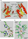
Structure and molecular mechanism of Bacillus anthracis cofactor-independent phosphoglycerate mutase: a crucial enzyme for spores and growing cells of Bacillus species
- PMID: 17085493
- PMCID: PMC1779985
- DOI: 10.1529/biophysj.106.093872
Structure and molecular mechanism of Bacillus anthracis cofactor-independent phosphoglycerate mutase: a crucial enzyme for spores and growing cells of Bacillus species
Abstract
Phosphoglycerate mutases (PGMs) catalyze the isomerization of 2- and 3-phosphoglycerates and are essential for glucose metabolism in most organisms. This study reports the production, structure, and molecular dynamics analysis of Bacillus anthracis cofactor-independent PGM (iPGM). The three-dimensional structure of B. anthracis PGM is composed of two structural and functional domains, the phosphatase and transferase. The structural relationship between these two domains is different than in the B. stearothermophilus iPGM structure determined previously. However, the structures of the two domains of B. anthracis iPGM show a high degree of similarity to those in B. stearothermophilus iPGM. The novel domain arrangement in B. anthracis iPGM and the dynamic property of these domains is directly linked to the mechanism of enzyme catalysis, in which substrate binding is proposed to result in close association of the two domains. The structure of B. anthracis iPGM and the molecular dynamics of this structure provide unique insight into the mechanism of iPGM catalysis, in particular the roles of changes in coordination geometry of the enzyme's two bivalent metal ions and the regulation of this enzyme's activity by changes in intracellular pH during spore formation and germination in Bacillus species.
Figures


Similar articles
-
Insights into the catalytic mechanism of cofactor-independent phosphoglycerate mutase from X-ray crystallography, simulated dynamics and molecular modeling.J Mol Biol. 2003 May 9;328(4):909-20. doi: 10.1016/s0022-2836(03)00350-4. J Mol Biol. 2003. PMID: 12729763
-
Mechanism of catalysis of the cofactor-independent phosphoglycerate mutase from Bacillus stearothermophilus. Crystal structure of the complex with 2-phosphoglycerate.J Biol Chem. 2000 Jul 28;275(30):23146-53. doi: 10.1074/jbc.M002544200. J Biol Chem. 2000. PMID: 10764795
-
Three-dimensional structure and molecular mechanism of novel enzymes of spore-forming bacteria.Med Sci Monit. 2002 Aug;8(8):RA183-90. Med Sci Monit. 2002. PMID: 12165756 Review.
-
Phosphorylation-mediated regulation of the Bacillus anthracis phosphoglycerate mutase by the Ser/Thr protein kinase PrkC.Biochem Biophys Res Commun. 2023 Jul 12;665:88-97. doi: 10.1016/j.bbrc.2023.04.039. Epub 2023 Apr 25. Biochem Biophys Res Commun. 2023. PMID: 37149987
-
Comparison of the binuclear metalloenzymes diphosphoglycerate-independent phosphoglycerate mutase and alkaline phosphatase: their mechanism of catalysis via a phosphoserine intermediate.Chem Rev. 2001 Mar;101(3):607-18. doi: 10.1021/cr000253a. Chem Rev. 2001. PMID: 11712498 Review. No abstract available.
Cited by
-
Identification of Genes Controlled by the Essential YycFG Two-Component System Reveals a Role for Biofilm Modulation in Staphylococcus epidermidis.Front Microbiol. 2017 Apr 26;8:724. doi: 10.3389/fmicb.2017.00724. eCollection 2017. Front Microbiol. 2017. PMID: 28491057 Free PMC article.
-
Unliganded structure of human bisphosphoglycerate mutase reveals side-chain movements induced by ligand binding.Acta Crystallogr Sect F Struct Biol Cryst Commun. 2010 Nov 1;66(Pt 11):1415-20. doi: 10.1107/S1744309110035475. Epub 2010 Oct 27. Acta Crystallogr Sect F Struct Biol Cryst Commun. 2010. PMID: 21045285 Free PMC article.
-
Exoproteomic analysis of two MLST clade 2 strains of Clostridioides difficile from Latin America reveal close similarities.Sci Rep. 2021 Jun 24;11(1):13273. doi: 10.1038/s41598-021-92684-0. Sci Rep. 2021. PMID: 34168208 Free PMC article.
-
Structural and Functional Characterization of the BcsG Subunit of the Cellulose Synthase in Salmonella typhimurium.J Mol Biol. 2018 Sep 14;430(18 Pt B):3170-3189. doi: 10.1016/j.jmb.2018.07.008. Epub 2018 Jul 12. J Mol Biol. 2018. PMID: 30017920 Free PMC article.
-
Macrocycle peptides delineate locked-open inhibition mechanism for microorganism phosphoglycerate mutases.Nat Commun. 2017 Apr 3;8:14932. doi: 10.1038/ncomms14932. Nat Commun. 2017. PMID: 28368002 Free PMC article.
References
-
- Jedrzejas, M. J. 2000. Structure, function, and evolution of phosphoglycerate mutases: comparison with fructose-2,6-bisphosphatase, acid phosphatase, and alkaline phosphatase. Prog. Biophys. Mol. Biol. 73:263–287. - PubMed
-
- Fothergill-Gilmore, L. A., and H. C. Watson. 1989. The phosphoglycerate mutases. Adv. Enzymol. Relat. Areas Mol. Biol. 62:227–313. - PubMed
-
- Setlow, P., and A. Kornberg. 1970. Biochemical studies of bacterial sporulation and germination. XXII. Energy metabolism in early stages of germination of Bacillus megaterium spores. J. Biol. Chem. 245:3637–3644. - PubMed
-
- Chander, M., B. Setlow, and P. Setlow. 1998. The enzymatic activity of phosphoglycerate mutase from gram-positive endospore-forming bacteria requires Mn2+ and is pH sensitive. Can. J. Microbiol. 44:759–767. - PubMed
-
- Kuhn, N. J., B. Setlow, and P. Setlow. 1993. Manganese(II) activation of 3-phosphoglycerate mutase of Bacillus megaterium: pH-sensitive interconversion of active and inactive forms. Arch. Biochem. Biophys. 306:342–349. - PubMed
Publication types
MeSH terms
Substances
Associated data
- Actions
LinkOut - more resources
Full Text Sources
Other Literature Sources
Molecular Biology Databases

