Knockout of the AtCESA2 gene affects microtubule orientation and causes abnormal cell expansion in Arabidopsis
- PMID: 17085513
- PMCID: PMC1761977
- DOI: 10.1104/pp.106.088393
Knockout of the AtCESA2 gene affects microtubule orientation and causes abnormal cell expansion in Arabidopsis
Abstract
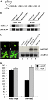
Complete cellulose synthesis is required to form functional cell walls and to facilitate proper cell expansion during plant growth. AtCESA2 is a member of the cellulose synthase A family in Arabidopsis (Arabidopsis thaliana) that participates in cell wall formation. By analysis of transgenic seedlings, we demonstrated that AtCESA2 was expressed in all organs, except root hairs. The atcesa2 mutant was devoid of AtCESA2 expression, leading to the stunted growth of hypocotyls in seedlings and greatly reduced seed production in mature plants. These observations were attributed to alterations in cell size as a result of reduced cellulose synthesis in the mutant. The orientation of microtubules was also altered in the atcesa2 mutant, which was clearly observed in hypocotyls and petioles. Complementary expression of AtCESA2 in atcesa2 could rescue the mutant phenotypes. Together, we conclude that disruption of cellulose synthesis results in altered orientation of microtubules and eventually leads to abnormal plant growth. We also demonstrated that the zinc finger-like domain of AtCESA2 could homodimerize, possibly contributing to rosette assemblies of cellulose synthase A within plasma membranes.
Figures


Similar articles
-
Cellulose Synthase Mutants Distinctively Affect Cell Growth and Cell Wall Integrity for Plant Biomass Production in Arabidopsis.Plant Cell Physiol. 2018 Jun 1;59(6):1144-1157. doi: 10.1093/pcp/pcy050. Plant Cell Physiol. 2018. PMID: 29514326
-
Wall architecture in the cellulose-deficient rsw1 mutant of Arabidopsis thaliana: microfibrils but not microtubules lose their transverse alignment before microfibrils become unrecognizable in the mitotic and elongation zones of roots.Protoplasma. 2001;215(1-4):172-83. doi: 10.1007/BF01280312. Protoplasma. 2001. PMID: 11732056
-
Anisotropic Cell Expansion Is Affected through the Bidirectional Mobility of Cellulose Synthase Complexes and Phosphorylation at Two Critical Residues on CESA3.Plant Physiol. 2016 May;171(1):242-50. doi: 10.1104/pp.15.01874. Epub 2016 Mar 11. Plant Physiol. 2016. PMID: 26969722 Free PMC article.
-
Cracking the elusive alignment hypothesis: the microtubule-cellulose synthase nexus unraveled.Trends Plant Sci. 2012 Nov;17(11):666-74. doi: 10.1016/j.tplants.2012.06.003. Epub 2012 Jul 9. Trends Plant Sci. 2012. PMID: 22784824 Free PMC article. Review.
-
PHS1ΔP as a promising tool to study microtubule-related processes in plant sciences.Plant Biol (Stuttg). 2025 Jun;27(4):445-449. doi: 10.1111/plb.70019. Epub 2025 Apr 1. Plant Biol (Stuttg). 2025. PMID: 40169153 Review.
Cited by
-
Cortical microtubule patterning in roots of Arabidopsis thaliana primary cell wall mutants reveals the bidirectional interplay with cell expansion.Plant Signal Behav. 2015;10(6):e1028701. doi: 10.1080/15592324.2015.1028701. Plant Signal Behav. 2015. PMID: 26042727 Free PMC article.
-
A kinetic analysis of the auxin transcriptome reveals cell wall remodeling proteins that modulate lateral root development in Arabidopsis.Plant Cell. 2013 Sep;25(9):3329-46. doi: 10.1105/tpc.113.114868. Epub 2013 Sep 17. Plant Cell. 2013. PMID: 24045021 Free PMC article.
-
Large-scale phosphoproteome analysis in seedling leaves of Brachypodium distachyon L.BMC Genomics. 2014 May 16;15(1):375. doi: 10.1186/1471-2164-15-375. BMC Genomics. 2014. PMID: 24885693 Free PMC article.
-
Identification and characterization of NARROW AND ROLLED LEAF 1, a novel gene regulating leaf morphology and plant architecture in rice.Plant Mol Biol. 2010 Jun;73(3):283-92. doi: 10.1007/s11103-010-9614-7. Epub 2010 Feb 13. Plant Mol Biol. 2010. PMID: 20155303
-
Physiology and Gene Expression Analysis of Tomato (Solanum lycopersicum L.) Exposed to Combined-Virus and Drought Stresses.Plant Pathol J. 2023 Oct;39(5):466-485. doi: 10.5423/PPJ.OA.07.2023.0103. Epub 2023 Oct 1. Plant Pathol J. 2023. PMID: 37817493 Free PMC article.
References
-
- Arioli T, Peng L, Betzner AS, Burn J, Wittke W, Herth W, Camilleri C, Hofte H, Plazinski J, Birch R, et al (1998) Molecular analysis of cellulose biosynthesis in Arabidopsis. Science 279 717–720 - PubMed
-
- Baskin TI (2001) On the alignment of cellulose microfibrils by cortical microtubules: a review and a model. Protoplasma 215 150–171 - PubMed
-
- Bent AF, Clough SJ (1998) Agrobacterium germ-line transformation: transformation of Arabidopsis without tissue culture. In SB Gelvin, RA Schilperoot, eds, Plant Molecular Biology Manual, Ed 2. Kluwer Academic Publishers, Dordrecht, The Netherlands, pp 1–14
Publication types
MeSH terms
Substances
LinkOut - more resources
Full Text Sources
Other Literature Sources
Molecular Biology Databases

