Review
. 2007 Jan;189(2):298-304.
doi: 10.1128/JB.01215-06. Epub 2006 Nov 3.

A new look at bacteriophage lambda genetic networks

Affiliations
Review

A new look at bacteriophage lambda genetic networks

Donald L Court et al. J Bacteriol. 2007 Jan.
No abstract available


Figures

FIG. 1.
FIG. 1.
Gene and transcription map of λ. Genes are shown in the shaded rectangle. The early transcripts for pL and pR promoters are shown as red arrows. The late transcript from pR′ is indicated with black arrows. The CII-activated pI, pRE, and pAQ transcripts are indicated with blue arrows. The pRM transcript activated by CI is a green arrow. Transcription terminators (t) are shown as red letters among the genes. The tI terminator is indicated in parenthesis because it is contained within the larger sib processing site. The operators OL and OR where CI and Cro bind are shown next to the pL and pR promoters.
FIG. 2.
FIG. 2.
Looping of the λ operators OL and OR during CI binding. The promoters pL and pR control the transcription of the early genes N and cIII and the genes cro and cII, respectively (Fig. 1). The promoter pRM transcribes CI, rexA, and rexB in a lysogen. The function of the rex genes in a lysogen is to exclude certain other phages infecting the cell. In the lower part of the figure, the repressor is shown binding to OL and OR regions to create a stable looped complex. At each individual operator, e.g., OL1, a dimer of repressor forms as indicated by the two dumbbells bound there. A similar dumbbell bound at OL2 forms a tetramer with the one at OL1, and together this tetramer interacts with a second tetramer at OR1-OR2 to form a looped octamer to tightly repress pL and pR. A repressor dimer is shown bound at OR3 interacting as a tetramer with another dimer at OL3. This OR3-OL3 tetramer represses pRM to down-regulate CI synthesis. As repressor levels drop in cells, OL3 and OR3 become free of repressor, and the pRM promoter is activated by repressor bound at OR2 (not shown).
FIG. 3.
FIG. 3.
RNase III control of N-mediated translation repression. RNA Pol is modified by N and Nus factors bound to the NutL RNA structure to become transcription termination resistant. This transcription antitermination complex represses N gene translation (top). If sufficient RNase III endonuclease is present, RNase III cleaves at RIII and dissociates the RNA Pol complex from the N gene-containing transcript, thereby preventing N translation repression. RTS denotes the DNA encoding the RIII RNase III structure, and SD is the N Shine-Dalgarno sequence.
FIG. 4.
FIG. 4.
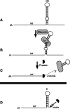
Regulation of int gene expression. (A) Int transcription from pL is antiterminated by N and reads through a terminator tI (see panel D) generating an extended stem structure (sib). (B) This structure is processed by RNase III to generate a new RNA 3′ end of int transcripts that is sensitive to the exonuclease PNPase, shown in panel C. (D) Transcription from pI is not antiterminated by N and stops at tI to generate a stem that is not processed by RNase III and that is resistant to PNPase.
FIG. 5.
FIG. 5.
l DNA integration and excision. The l DNA molecule circularizes following infection (top), and with Int protein of the phage and IHF protein of E. coli, recombines at specific sites attP-P′ in the phage and attB-B′ in the bacterial chromosome (shown as a linear shaded bar) to integrate. The integration event generates hybrid att sites attB-P′ and attP-B′ at the prophage DNA (black bar) ends. Following prophage induction, excision occurs between attB-P′ and attP-B′ by site-specific recombination. For this recombination the Xis protein of the phage is required in addition to Int and IHF. An important rearrangement of the phage DNA is caused by the integration step. The sib site is adjacent to int during the phage infection (top). However, integration completely separates sib from int (bottom) in the prophage. Thus, integration changes the regulation of Int expression from pL transcripts.

References

    1. Atsumi, S., and J. W. Little. 2004. Regulatory circuit design and evolution using phage lambda. Genes Dev. 18:2086-2094. - PMC - PubMed
    1. Atsumi, S., and J. W. Little. 2006. Role of the lytic repressor in prophage induction of phage λ as analyzed by a module-replacement approach. Proc. Natl. Acad. Sci. USA. 103:4558-4563. - PMC - PubMed
    1. Aurell, E., S. Brown, J. Johanson, and K. Sneppen. 2002. Stability puzzles in phage lambda. Phys. Rev. E 65:051914-051919. - PubMed
    1. Banuett, F., M. A. Hoyt, L. McFarlane, H. Echols, and I. Herskowitz. 1986. hflB, a new Escherichia coli locus regulating lysogeny and the level of bacteriophage lambda cII protein. J. Mol. Biol. 187:213-224. - PubMed
    1. Britton, R. A., B. S. Powell, S. Dasgupta, Q. Sun, W. Margolin, J. R. Lupski, and D. L. Court. 1998. Cell cycle arrest in Era GTPase mutants: a potential growth rate-regulated checkpoint in Escherichia coli. Mol. Microbiol. 27:739-750. - PubMed

Publication types

LinkOut - more resources