. 2007 Jan;189(2):305-12.
doi: 10.1128/JB.01281-06. Epub 2006 Nov 3.

Mutational analysis of the flagellar protein FliG: sites of interaction with FliM and implications for organization of the switch complex

Affiliations

Mutational analysis of the flagellar protein FliG: sites of interaction with FliM and implications for organization of the switch complex

Perry N Brown et al. J Bacteriol. 2007 Jan.

Abstract

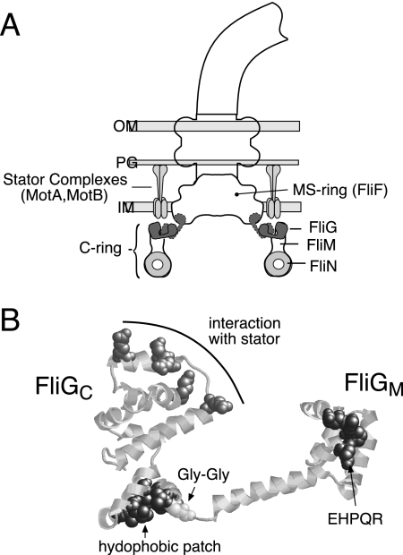
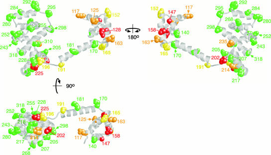
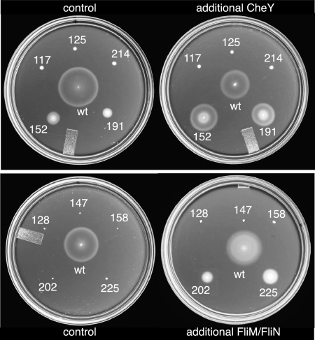
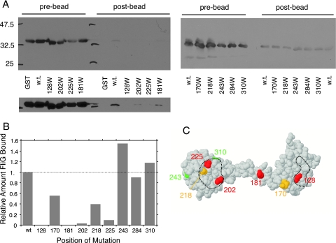
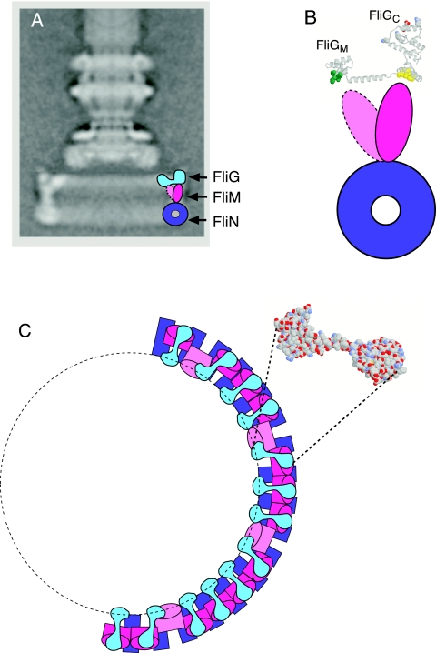
The switch complex at the base of the bacterial flagellum is essential for flagellar assembly, rotation, and switching. In Escherichia coli and Salmonella, the complex contains about 26 copies of FliG, 34 copies of FliM, and more then 100 copies of FliN, together forming the basal body C ring. FliG is involved most directly in motor rotation and is located in the upper (membrane-proximal) part of the C ring. A crystal structure of the middle and C-terminal parts of FliG shows two globular domains connected by an alpha-helix and a short extended segment. The middle domain of FliG has a conserved surface patch formed by the residues EHPQ(125-128) and R(160) (the EHPQR motif), and the C-terminal domain has a conserved surface hydrophobic patch. To examine the functional importance of these and other surface features of FliG, we made mutations in residues distributed over the protein surface and measured the effects on flagellar assembly and function. Mutations preventing flagellar assembly occurred mainly in the vicinity of the EHPQR motif and the hydrophobic patch. Mutations causing aberrant clockwise or counterclockwise motor bias occurred in these same regions and in the waist between the upper and lower parts of the C-terminal domain. Pull-down assays with glutathione S-transferase-FliM showed that FliG interacts with FliM through both the EHPQR motif and the hydrophobic patch. We propose a model for the organization of FliG and FliM subunits that accounts for the FliG-FliM interactions identified here and for the different copy numbers of FliG and FliM in the flagellum.


Figures

FIG. 1.
FIG. 1.
(A) Locations of proteins involved in flagellar rotation. The location of FliN is deduced from targeted cross-linking studies (39) and electron microscopic reconstructions (12, 48, 50, 58). Although FliG is known to be in the upper part of the C ring (16, 22, 26, 37, 61) and some features of its organization have been deduced from cross-linking (31), its exact location is not yet certain. Accordingly, two possible locations for FliG are indicated. OM, outer membrane; PG, peptidoglycan; IM, inner membrane. (B) Structure of residues 115 to 327 of T. maritima FliG (FliGMC) (7), highlighting conserved surface features.
FIG. 2.
FIG. 2.
Locations of the Trp replacement mutations on FliGMC and their swarming phenotypes. Green, swarming rate (relative to wild type) of 0.7 or better; yellow, relative rate of between 0.1 and 0.3; orange, rate nonzero but less than 0.1; red, nonswarming.
FIG. 3.
FIG. 3.
Enhancement of swarming by overexpression of CheY (top) or FliM and FliN (bottom) in selected FliG mutants. CheY or FliM-FliN were expressed from plasmids, as described in Materials and Methods. Control strains contained a second plasmid that encoded the relevant antibiotic resistance but no flagellar genes. Swarms were allowed to develop for approximately 9 h for all of the mutants and for 5 h for the wild type (wt).
FIG. 4.
FIG. 4.
(A) Coisolation of FliG with GST-FliM and effects of FliG mutations on the binding. Coisolated FliG was detected using anti-FliG immunoblots. Prebead controls show the relative levels of FliG present in each sample prior to treatment with the beads, and postbead samples show levels of FliG coisolated with GST-FliM. Positions of the Trp mutations are shown at the bottom. Some of the mutations weakened binding so much that coisolated FliG could be seen only in a longer exposure, shown at the bottom for the left-hand gel. w.t., wild type. (B) Relative levels of coisolated wild-type and mutant FliG proteins. Results of a typical binding experiment are shown. (C) Mutations mapped onto the FliG structure. Red, binding reduced to 10% or less of wild-type level; orange, binding reduced to about half of wild-type level; green, binding similar to wild-type level. Dashed lines indicate the hydrophobic patch (left) and EHPQR motif (right). Position 284 (which did not affect the binding to FliM and so would be green) is not visible in this view but is on the charge-bearing ridge of the C-terminal domain (Fig. 2).
FIG. 5.
FIG. 5.
Model for subunit organization in the switch complex. (A) Proposed locations of FliG, FliM, and FliN in the C ring. FliG is placed in the more outboard of the two locations shown in Fig. 1, to allow interaction between FliM and the middle domain of FliG (the EHPQR motif). (B) Enlarged view of the hypothesized FliG-FliM interactions. FliM is pictured in two orientations, one interacting with the EHPQR motif in the FliG middle domain (green) and the other interacting with the hydrophobic patch in the FliG C-terminal domain (yellow). (C) View from the “top” (the membrane-proximal side) of the C ring. Subunit organization is illustrated for half of the ring, using the same coloring as in panel A. The shape of the FliG subunits is based on the FliG structure, with the relative orientations of the domains adjusted slightly to account for targeted cross-linking results reported previously (31). The structure of a FliG subunit, viewed from membrane-proximal side, is shown for comparison. In the model, 26 copies of FliM attach to the C-terminal domain of FliG through the hydrophobic patch, while the approximately eight “extra” FliM subunits tilt inward to interact with the middle domain of FliG.

Comment in

References

    1. Attmannspacher, U., B. Scharf, and R. Schmitt. 2005. Control of speed modulation (chemokinesis) in the unidirectional rotary motor of Sinorhizobium meliloti. Mol. Microbiol. 56:708-718. - PubMed
    1. Berg, H. C. 2003. The rotary motor of bacterial flagella. Annu. Rev. Biochem. 72:19-54. - PubMed
    1. Berry, R. M., and H. C. Berg. 1999. Torque generated by the flagellar motor of Escherichia coli while driven backward. Biophys. J. 76:580-587. - PMC - PubMed
    1. Blair, D. F., and H. C. Berg. 1988. Restoration of torque in defective flagellar motors. Science 242:1678-1681. - PubMed
    1. Block, S. M., and H. C. Berg. 1984. Successive incorporation of force-generating units in the bacterial rotary motor. Nature 309:470-472. - PubMed

Publication types

MeSH terms