

A pivotal role for endogenous TGF-beta-activated kinase-1 in the LKB1/AMP-activated protein kinase energy-sensor pathway
- PMID: 17085580
- PMCID: PMC1859937
- DOI: 10.1073/pnas.0604708103
A pivotal role for endogenous TGF-beta-activated kinase-1 in the LKB1/AMP-activated protein kinase energy-sensor pathway
Abstract
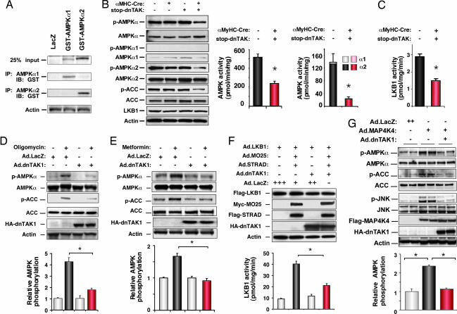
TGF-beta-activated kinase-1 (TAK1), also known as MAPKK kinase-7 (MAP3K7), is a candidate effector of multiple circuits in cardiac biology and disease. Here, we show that inhibition of TAK1 in mice by a cardiac-specific dominant-negative mutation evokes electrophysiological and biochemical properties reminiscent of human Wolff-Parkinson-White syndrome, arising from mutations in AMP-activated protein kinase (AMPK), most notably, accelerated atrioventricular conduction and impaired AMPK activation. To test conclusively the biochemical connection from TAK1 to AMPK suggested by this phenotype, we disrupted TAK1 in mouse embryos and embryonic fibroblasts by Cre-mediated recombination. In TAK1-null embryos, the activating phosphorylation of AMPK at T172 was blocked, accompanied by defective AMPK activity. However, loss of endogenous TAK1 causes midgestation lethality, with defective yolk sac and intraembryonic vasculature. To preclude confounding lethal defects, we acutely ablated floxed TAK1 in culture by viral delivery of Cre. In culture, endogenous TAK1 was activated by oligomycin, the antidiabetic drug metformin, 5-aminoimidazole-4-carboxamide riboside (AICAR), and ischemia, well established triggers of AMPK activity. Loss of TAK1 in culture blocked T172 phosphorylation induced by all three agents, interfered with AMPK activation, impaired phosphorylation of the endogenous AMPK substrate acetyl CoA carboxylase, and also interfered with activation of the AMPK kinase LKB1. Thus, by disrupting the endogenous TAK1 locus, we prove a pivotal role for TAK1 in the LKB1/AMPK signaling axis, an essential governor of cell metabolism.
Conflict of interest statement
The authors declare no conflict of interest.
Figures


References
-
- Johnson GL, Lapadat R. Science. 2002;298:1911–1912. - PubMed
-
- Yamaguchi K, Shirakabe K, Shibuya H, Irie K, Oishi I, Ueno N, Taniguchi T, Nishida E, Matsumoto K. Science. 1995;270:2008–2011. - PubMed
-
- Jadrich JL, O'Connor MB, Coucouvanis E. Development (Cambridge, UK) 2006;133:1529–1541. - PubMed
-
- Branda CS, Dymecki SM. Dev Cell. 2004;6:7–28. - PubMed
Publication types
MeSH terms
Substances
LinkOut - more resources
Full Text Sources
Molecular Biology Databases
Research Materials
Miscellaneous
