. 2006 Nov 14;103(46):17225-30.
doi: 10.1073/pnas.0607171103. Epub 2006 Nov 3.

Insights into transcription enhancer factor 1 (TEF-1) activity from the solution structure of the TEA domain

Affiliations

Insights into transcription enhancer factor 1 (TEF-1) activity from the solution structure of the TEA domain

Asokan Anbanandam et al. Proc Natl Acad Sci U S A. .

Abstract

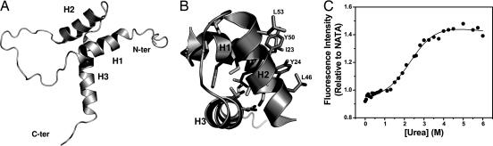
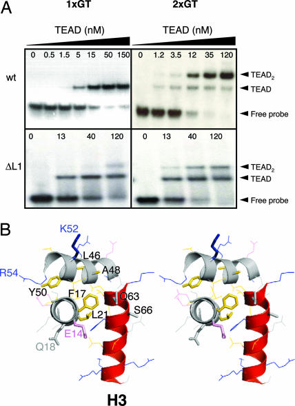
Transcription enhancer factor 1 is essential for cardiac, skeletal, and smooth muscle development and uses its N-terminal TEA domain (TEAD) to bind M-CAT elements. Here, we present the first structure of TEAD and show that it is a three-helix bundle with a homeodomain fold. Structural data reveal how TEAD binds DNA. Using structure-function correlations, we find that the L1 loop is essential for cooperative loading of TEAD molecules on to tandemly duplicated M-CAT sites. Furthermore, using a microarray chip-based assay, we establish that known binding sites of the full-length protein are only a subset of DNA elements recognized by TEAD. Our results provide a model for understanding the regulation of genome-wide gene expression during development by TEA/ATTS family of transcription factors.


Conflict of interest statement

The authors declare no conflict of interest.

Figures

Fig. 1.
Fig. 1.
TEAD transcription factors. (A) TEAD proteins regulate tissue-development in eukaryotes. TEF-1 also regulates viral gene activation and TEC1 homolog is essential for Candida virulence. (B) TEAD sequence alignment shows strict evolutionary conservation of most residues (white letters on black background). TEAD used here contains five extraneous residues (in italics) resulting from cloning procedures. Residue 2 of our TEAD corresponds to residue 28 of the TEF-1. Positions of secondary structural elements, based on our NMR-derived structure, are shown over the sequence. Mutations discussed in the article are identified by ▾.
Fig. 2.
Fig. 2.
Solution NMR structure of TEAD. (A) Ribbon diagram shows front view of the TEAD fold in which helices H1 and H2 pack against H3. N and C termini are labeled. (B) View down the H3 helix illustrates hydrophobic residues (sticks) contributing to core packing and the surface hydrophobic patch. (C) TEAD unfolds with a midpoint of 2.5 M urea. Fluorescence intensity is relative to that of an equimolar solution of N-acetyl tryptophanamide (NATA).
Fig. 3.
Fig. 3.
DNA-binding activity of TEAD. (A) EMSA. (B) Nonlinear least-squares fit (solid line) of EMSA data (circles) and 95% confidence limits (dotted lines). (C) Design of DNA sequences used in D-LIA. DNA is attached to the chip by using a linker containing thymidines (T) and poly(ethylene glycol) (P). N5 = GCTATG or GGGGG. (D) Fluorescent spots extracted from the inverted fluorescence image of a microfluidic chip show TEAD binding to various synthetic double-stranded DNA elements. Dark and light spots indicate strong and weak binding, respectively. ∗, DNA sequence is given in Supporting Text. (E) Identification of DNA length suitable for NMR investigations of protein–DNA complex.
Fig. 4.
Fig. 4.
NMR-mapping of DNA-binding surface. (A) 1H,15N-HSQC spectra of DNA-free (black) and DNA-bound (red) TEAD. Labeled residues show unambiguous and substantial chemical shift changes upon binding DNA. Amino acids in the L1 loop are shown in italics. Asterisks denote side-chain resonances. (B) Backbone (orange) and side-chain (red) resonances that respond to DNA binding identify the DNA-binding surface. (Left) Front view. (Right) Back view. (C) Surface electrostatic potential of TEAD. The front surface (Left) comprises some negatively charged (red) residues, whereas back surface (Right) lacks negative charges.
Fig. 5.
Fig. 5.
Models of TEAD structure when bound to DNA. (A) The MatA1 homeodomain (gold) in the MatA1/Matα2/DNA complex (PDB ID 1AKH; ref. 62) is the closest structural homolog of TEAD. (B) Model of TEAD/DNA complex generated by superimposition of H2 and H3 helices of TEAD on MatA1.
Fig. 6.
Fig. 6.
Correlating TEAD structure and function. (A) EMSA of wild-type and mutant TEAD. Wild-type TEAD molecules bind cooperatively to tandem M-CAT-like elements (2xGT). The L1 deletion mutant of TEAD binds M-CAT DNA stochastically, and cooperativity is lost. (B) Stereoview of TEAD structure is shown. Amino acid side chains in the three helices are shown as lines or sticks (mutants are also labeled). Loops are not shown for clarity of illustration. Gly-28 resides in loop L1. DNA-recognition helix, H3, is shown in red. Side-chain colors: pink, acidic; blue, basic; gold, hydrophobic; gray, other.
Fig. 7.
Fig. 7.
SRF and TEF-1 DNA-binding domains do not load on to adjacent elements cooperatively.

References

    1. Davidson I, Xiao JH, Rosales R, Staub A, Chambon P. Cell. 1988;54:931–942. - PubMed
    1. Ishiji T, Lace MJ, Parkkinen S, Anderson RD, Haugen TH, Cripe TP, Xiao JH, Davidson I, Chambon P, Turek LP. EMBO J. 1992;11:2271–2281. - PMC - PubMed
    1. Mar JH, Ordahl CP. Mol Cell Biol. 1990;10:4271–4283. - PMC - PubMed
    1. Xiao JH, Davidson I, Matthes H, Garnier JM, Chambon P. Cell. 1991;65:551–568. - PubMed
    1. Campbell S, Inamdar M, Rodrigues V, Raghavan V, Palazzolo M, Chovnick A. Genes Dev. 1992;6:367–379. - PubMed

Publication types

Associated data

LinkOut - more resources