

Structural basis for protein-protein interactions in the 14-3-3 protein family
- PMID: 17085597
- PMCID: PMC1859916
- DOI: 10.1073/pnas.0605779103
Structural basis for protein-protein interactions in the 14-3-3 protein family
Abstract
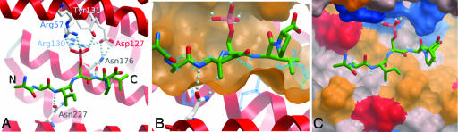
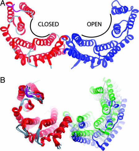
The seven members of the human 14-3-3 protein family regulate a diverse range of cell signaling pathways by formation of protein-protein complexes with signaling proteins that contain phosphorylated Ser/Thr residues within specific sequence motifs. Previously, crystal structures of three 14-3-3 isoforms (zeta, sigma, and tau) have been reported, with structural data for two isoforms deposited in the Protein Data Bank (zeta and sigma). In this study, we provide structural detail for five 14-3-3 isoforms bound to ligands, providing structural coverage for all isoforms of a human protein family. A comparative structural analysis of the seven 14-3-3 proteins revealed specificity determinants for binding of phosphopeptides in a specific orientation, target domain interaction surfaces and flexible adaptation of 14-3-3 proteins through domain movements. Specifically, the structures of the beta isoform in its apo and peptide bound forms showed that its binding site can exhibit structural flexibility to facilitate binding of its protein and peptide partners. In addition, the complex of 14-3-3 beta with the exoenzyme S peptide displayed a secondary structural element in the 14-3-3 peptide binding groove. These results show that the 14-3-3 proteins are adaptable structures in which internal flexibility is likely to facilitate recognition and binding of their interaction partners.
Conflict of interest statement
The authors declare no conflict of interest.
Figures


References
-
- Moore BW, Perez VJ. Specific Acidic Proteins of the Nervous System. Englewood Cliffs, NJ: Prentice Hall; 1967.
-
- Chaudhri M, Scarabel M, Aitken A. Biochem Biophys Res Commun. 2003;300:679–685. - PubMed
-
- Jones DH, Ley S, Aitken A. FEBS Lett. 1995;368:55–58. - PubMed
-
- Jin J, Smith FD, Stark C, Wells CD, Fawcett JP, Kulkarni S, Metalnikov P, O'Donnell P, Taylor P, Taylor L, et al. Curr Biol. 2004;14:1436–1450. - PubMed
-
- Darling DL, Yingling J, Wynshaw-Boris A. Curr Top Dev Biol. 2005;68:281–315. - PubMed
Publication types
MeSH terms
Substances
Associated data
- Actions
- Actions
- Actions
- Actions
- Actions
- Actions
- Actions
Grants and funding
LinkOut - more resources
Full Text Sources
Molecular Biology Databases
