. 2007 Jan;73(2):586-93.
doi: 10.1128/AEM.01839-06. Epub 2006 Nov 3.

Poly(3-hydroxybutyrate) granules at the early stages of formation are localized close to the cytoplasmic membrane in Caryophanon latum

Affiliations

Poly(3-hydroxybutyrate) granules at the early stages of formation are localized close to the cytoplasmic membrane in Caryophanon latum

Dieter Jendrossek et al. Appl Environ Microbiol. 2007 Jan.

Abstract

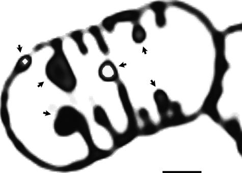
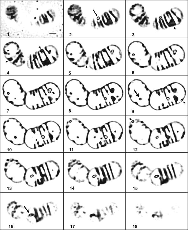
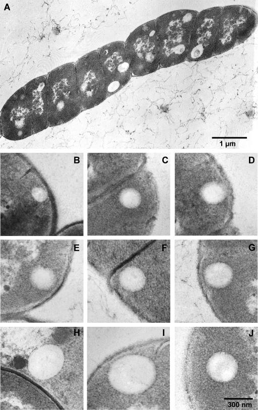
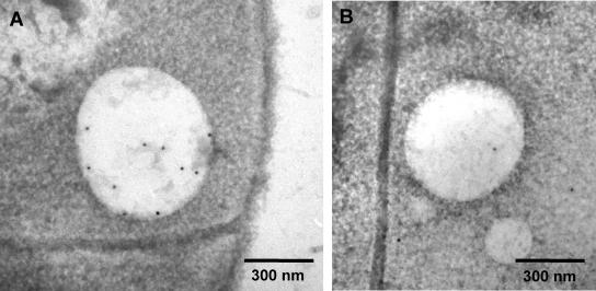
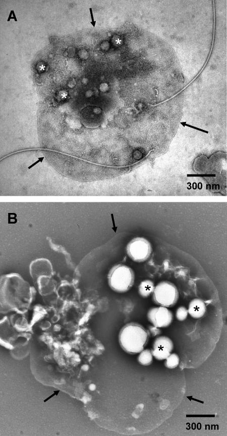
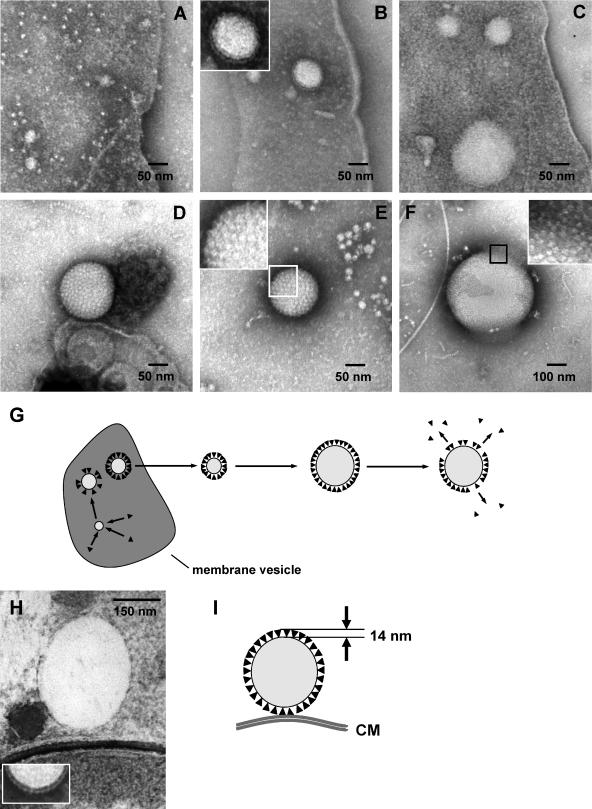
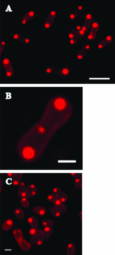
Localization of newly synthesized poly(3hydroxybutyrate) (PHB) granules was determined by confocal laser scanning fluorescence microscopy of Nile red-stained cells and by transmission electron microscopy (TEM). PHB granules of Nile red-stained living cells of Caryophanon latum at the early stages of PHB accumulation were frequently found at or close to the cytoplasmic membrane. TEM analysis of the same culture revealed electron-translucent globular structures resembling PHB granules that were nonrandomly distributed in the cell lumen but were frequently found at or close to the cytoplasmic membrane. Immunogold labeling using PHB-specific antiserum confirmed that the electron-translucent structures represented PHB granules. Electron microscopy examination of PHB granules after cell lysis revealed that PHB granules were often associated with membrane vesicles. Nonrandom localization of PHB granules was also found in Beijerinckia indica. Cells of this species harbored one PHB granule at each cell pole. Our results show that newly synthesized PHB granules often are close to or even in physical contact with the cytoplasmic membrane. Possible explanations for this unexpected finding and a hypothetical model of PHB granule formation in C. latum are discussed.


Figures

FIG. 1.
FIG. 1.
Confocal laser scanning fluorescence microscopy of C. latum. Shown is a typical image of PHB granules in a C. latum trichome after Nile red staining (red color in gray). The granules were exclusively found close to the membrane of the bacterium (arrowheads). The image shows a volume rendering of three optical sections with 120-nm steps each, representing a slice of about 360 nm. Color levels above 200 (of the 255 levels of the 8-bit image) have been set to 255 to mark the granules (white). The scale bar represents 1 μm.
FIG. 2.
FIG. 2.
Confocal laser scanning fluorescence microscopy of C. latum. Shown is a series of optical sections through the same trichome as in Fig. 1. Each image is a volume rendering of two optical sections with a 120-nm step size, hence representing about 240 nm of the organism. The sections cover the whole volume of the cells. Color levels (in gray) above 200 (of the 255 levels of the 8-bit image) have been artificially stained in gray to mark the granules. The scale bar represents 1 μm. Arrowheads indicate PHB granules at or close to the cytoplasmic membrane. Arrows in slices 2 and 16 (thin arrows) indicate structures that are located close to the top or bottom layer of the cells. The big arrow in slice 9 indicates a PHB granule clearly detached from the membrane.
FIG. 3.
FIG. 3.
TEM analysis of C. latum: ultrathin sections of C. latum grown for 80 min on acetate medium. (A) Overview of a C. latum trichome. (B to J) Granules in immediate vicinity of the cytoplasmic membrane (B to I) and detached from the membrane (J). Note that the thickness of the PHB granule phasin layer is about 14 nm and that most PHB granules are not cut in the middle of the granule where the diameter would be maximal.
FIG. 4.
FIG. 4.
Immunolocalization of PHB in C. latum. (A) Anti-PHB antiserum. (B) PBS instead of antiserum (negative control).
FIG. 5.
FIG. 5.
TEM analysis of C. latum: membrane vesicles in culture samples after negative staining. Some of the granules are marked by asterisks. Arrows point to the membrane boundaries of the vesicles. (A) Sample taken from an overnight culture grown on acetate-free medium. (B) Sample taken from a culture grown on acetate medium for 7 h.
FIG. 6.
FIG. 6.
Proposed model of formation and structure of granules. (A to F) Granules (C. latum) at different stages of development. Shown are PHB granules attached to membrane vesicles (A to C) and free PHB granules (D to F). Cells were grown on acetate-free medium (A) or on acetate medium for 20 min (B and C) or for 16 h (D to F). The inset in panel B indicates 14-nm particles on the surface of the enlarged PHB granule. The insets in panels E and F show enlarged views of the boxed areas. (G) Schematic drawing of granule development as implied by the observations described in panels A to F. An ultrathin section (see Fig. 3H) is compared with a negatively stained sample (H). The distance between a granule and the cytoplasmic membrane may be easily explained by the particle shell measuring 14 nm in thickness (I).
FIG. 7.
FIG. 7.
Confocal laser scanning fluorescence microscopy of B. indica. Maximum projections of confocal stacks. A typical picture during growth is shown (24 h, overview) in panel A. The scale bar is 4 μm. Enlargement of one dumbbell-like cell harboring PHB granules at the two cell poles and one small PHB granule at the site of septum of the upcoming cell division is shown in panel B. The scale bar is 1 μm. Resting cells (48 h) became ellipsoid and often harbor only one PHB granule (C). The scale bar is 1 μm.

References

    1. Abe, T., T. Kobayashi, and T. Saito. 2005. Properties of a novel intracellular poly(3-hydroxybutyrate) depolymerase with high specific activity (PhaZd) in Wautersia eutropha H16. J. Bacteriol. 187:6982-6990. - PMC - PubMed
    1. Anderson, A. J., and E. A. Dawes. 1990. Occurrence, metabolism, metabolic role, and industrial uses of bacterial polyhydroxyalkanoates. Microbiol. Rev. 54:450-472. - PMC - PubMed
    1. Becking, J. 1992. The genus Beijerinckia, p. 2254-2267. In A. Balows, H. Trüper, M. Dworkin, W. Harder, and K. Schleifer (ed.), The prokaryotes, vol. III. Springer Verlag, New York, NY.
    1. Boatman, E. S. 1964. Observations on the fine structure of spheroplasts of Rhodospirillum rubrum. J. Cell Biol. 20:297-311. - PMC - PubMed
    1. Degelau, A., T. Scheper, J. Bailey, and C. Guske. 1995. Fluorometric measurements of poly-beta-hydroxybutyrate in Alcaligenes eutrophus by flow cytometry and spectrofluoromety. Appl. Microbiol. Biotechnol. 42:653-657.

Publication types

MeSH terms

LinkOut - more resources