. 2006 Dec;7(12):1326-33.
doi: 10.1038/ni1407. Epub 2006 Nov 5.

Integrin signaling in neutrophils and macrophages uses adaptors containing immunoreceptor tyrosine-based activation motifs

Affiliations

Integrin signaling in neutrophils and macrophages uses adaptors containing immunoreceptor tyrosine-based activation motifs

Attila Mócsai et al. Nat Immunol. 2006 Dec.

Abstract

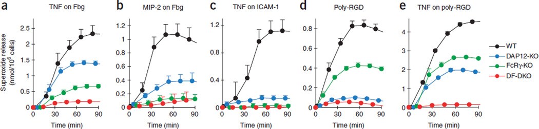
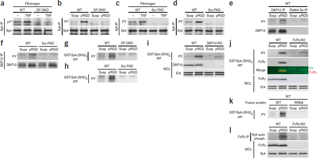
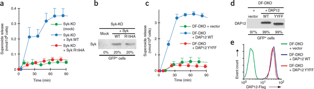
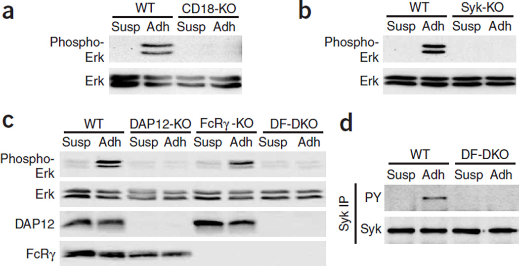
At sites of inflammation, ligation of leukocyte integrins is critical for the activation of cellular effector functions required for host defense. However, the signaling pathways linking integrin ligation to cellular responses are poorly understood. Here we show that integrin signaling in neutrophils and macrophages requires adaptors containing immunoreceptor tyrosine-based activation motifs (ITAMs). Neutrophils and macrophages lacking two ITAM-containing adaptor proteins, DAP12 and FcRgamma, were defective in integrin-mediated responses. Activation of the tyrosine kinase Syk by integrins required that DAP12 and FcRgamma were first phosphorylated by Src family kinases. Retroviral transduction of neutrophils and macrophages with wild-type and mutant Syk or DAP12 demonstrated that the Src homology 2 domains of Syk and the ITAM of DAP12 were required for integrin signaling. Our data show that integrin signaling for the activation of cellular responses in neutrophils and macrophages proceeds by an immunoreceptor-like mechanism.


Figures

Figure 1
Figure 1
Defective integrin-mediated respiratory burst in DF double-knockout neutrophils. Superoxide release of wild-type neutrophils (WT), DAP12-knockout neutrophils (DAP12-KO), FcRγ-knockout neutrophils (FcRγ-KO) and DF double-knockout neutrophils (DF-DKO) plated on surfaces coated with integrin ligand (fibrinogen (Fbg; a,b), ICAM-1 (c) or poly-RGD (d,e)) in the presence or absence of an additional stimulus (50 ng/ml of mouse TNF (a,c,e), 100 ng/ml of mouse MIP-2 (b) or none (d)). Unstimulated values (no TNF or MIP-2) were subtracted from stimulated values in a–c,e; error bars, s.d. of triplicate measurements. Data are representative of a minimum of three independent experiments each.
Figure 2
Figure 2
Defective integrin-mediated nonoxidative responses in DF double-knockout neutrophils. (a) Release of gelatinase granules by wild-type, DAP12-knockout, FcRγ-knockout and DF double-knockout neutrophils plated for 60 min on fibrinogen in the presence (TNF) or absence (− or Control) of 50 ng/ml of mouse TNF and analyzed by gelatinase zymogram. (b) Phase-contrast microscopy of wild-type and DF double-knockout neutrophils plated for 30 min on fibrinogen in the presence (right) or absence (left) of 50 ng/ml of mouse TNF. Original magnification, ×40. (c) Firm adherence of neutrophils treated as described in a. (d) Migration of wild-type and DF double-knockout neutrophils through FCS-coated Transwells in response to increasing concentrations of fMLP, assessed after 60 min. Data are representative of a minimum of three independent experiments each (error bars, s.d. of triplicate measurements).
Figure 3
Figure 3
Defective integrin-mediated signaling in DF double-knockout neutrophils. Immunoassays of lysates of wild-type and DF double-knockout neutrophils kept in suspension (Susp) or plated for 15 min on a surface coated with poly-RGD (pRGD). (a) Immunoblot analysis of total lysates with antibody to phosphorylated tyrosine (PY). (b,c) Immunoprecipitation (IP) with anti-Vav (b) or anti-Pyk2 (c) followed by immunoblot analysis (antibodies, left margin). (d,e) Immunoblot analysis of total lysates with antibody to phosphorylated Erk (Phospho-Erk) or to total Erk (Erk; d) or with antibody to phosphorylated p38 (Phospho-p38) or to total p38 (p38; e). All data are representative of three or more independent experiments.
Figure 4
Figure 4
Normal adhesion-independent responses of DF double-knockout neutrophils. (a,b) Flow cytometry of surface expression of CD11b (a) and CD18 (b) by wild-type or DF double-knockout neutrophils stimulated in suspension for 30 min with 50 ng/ml of mouse TNF or left unstimulated (Control). (c,d) Immunoblot of lysates of suspended wild-type or DF double-knockout neutrophils left unstimulated (−) or stimulated for 5 min with 50 ng/ml of mouse TNF (c) or for 10 min with 1 µg/ml of Pam3CSK4 (Pam3; d). Antibodies, left margin (Phospho-, phosphorylated). (e) Actin polymerization by suspended wild-type or DF double-knockout neutrophils stimulated with 10 ng/ml of mouse MIP-2. MFI, mean fluorescent intensity. (f,g) Release of intracellular calcium ([Ca2+]i) from suspended wild-type and DF double-knockout neutrophils stimulated with 100 ng/ml of mouse MIP-2 (f) or 3 µM fMLP (g). (h,i) Superoxide release from suspended wild-type and DF double-knockout neutrophils pretreated with 10 µM cytochalasin B and stimulated with 3 µM fMLP (h) or stimulated with 100 nM phorbol 12-myristate 13-acetate (PMA; i). Unstimulated values are subtracted in h and i. Data are representative of a minimum of three independent experiments each (error bars, s.d. of triplicate measurements).
Figure 5
Figure 5
Evidence of a Src family kinase–DAP12 or FcRγ–Syk pathway during integrin signaling of neutrophils. (a–d) Immunoassay of lysates of neutrophils plated on fibrinogen in the presence (TNF) or absence (−) of 50 ng/ml of mouse TNF (a,c) or on a surface coated with poly-RGD (b,d). Lysates prepared after 15 min of stimulation were immunoprecipitated with anti-Syk. Control, unstimulated cells in suspension (Susp; b,d). (e–l) Immunoassay of lysates of neutrophils plated on a surface coated with poly-RGD with (i,j,l) or without (e–h,k) 50 ng/ml of mouse TNF. Lysates prepared after 15 min of stimulation were immunoprecipitated with anti-DAP12 (e,f) or anti-FcRγ (l) or were precipitated (ppt) by incubation with GST-Syk-(SH2)2 (g–k). Control, unstimulated cells in suspension (Susp). (j) Merge, overlay of phosphorylated-tyrosine and FcRγ blots above. (l) Top, autophosphorylation of Syk (Syk auto-phosph.) in anti-FcRγ immunoprecipitates. Immunoblots are in reducing conditions except for e–h,k. Src-FKO, ‘Src family knockout’ (lacking Hck, Fgr and Lyn); Rabbit Se, normal rabbit serum; WCL, whole-cell lysate; RRAA, GST-Syk-(SH2)2 with R41A and R194A substitutions. All data are representative of three or more independent experiments.
Figure 6
Figure 6
Critical function for the Syk SH2 domains and the ITAM tyrosine residues of DAP12 for integrin-mediated responses of neutrophils. Retrovirus-transduced Syk-knockout (a,b) or DF double-knockout (c–e) fetal liver hematopoietic stem cells were injected into lethally irradiated recipient mice and, after reconstitution of the hematopoietic system, neutrophils were isolated for functional and gene expression studies. (a,c) Respiratory burst of neutrophils (purified from reconstituted mice) stimulated with 50 ng/ml of mouse TNF on a surface coated with poly-RGD (unstimulated values are subtracted; error bars, s.d. of triplicate measurements). (b,d) Immunoblot of protein expression and flow cytometry for percent GFP+ cells (below lanes). Control, neutrophils purified from mice reconstituted with stem cells that were ‘mock infected’ (Mock) or were infected with vector only (+ vector). (e) Flow cytometry for surface expression of exogenous DAP12 on transduced DF double-knockout neutrophils, detected by anti-Flag. YYFF, DAP12 Y65F and Y76F mutant. Data in a,b and in c–e are from the same experiment and are representative of three to five independent experiments.
Figure 7
Figure 7
Defective integrin-mediated responses in DF double-knockout macrophages. Bone marrow–derived macrophages were plated on Valmark dishes for 30–45 min (Adh) or were kept in suspension (Susp), then lysates were prepared. (a–c) Immunoblot analysis of Erk phosphorylation. (d) Immunoprecipitation with anti-Syk, followed by immunoblot analysis of tyrosine phosphorylation. All data are representative of three or more independent experiments.
Figure 8
Figure 8
Immunoreceptor-like signaling by macrophage integrins. (a,b) Immunoblot analysis of Erk phosphorylation and flow cytometry of percent GFP+ cells (below lanes) for Syk-knockout (a) or DF double-knockout (b) bone marrow–derived macrophages infected with retrovirus containing wild-type or mutant Syk or DAP12, respectively, then plated on Valmark dishes for 30–45 min; cell lysates were prepared and analyzed. (c) Flow cytometry for surface expression of exogenous DAP12 on GFP+ macrophages from a,b, detected by anti-Flag staining. Data are representative of at least four independent experiments.

Comment in

References

    1. Berton G, Lowell CA. Integrin signalling in neutrophils and macrophages. Cell. Signal. 1999;11:621–635. - PubMed
    1. Bunting M, Harris ES, McIntyre TM, Prescott SM, Zimmerman GA. Leukocyte adhesion deficiency syndromes: adhesion and tethering defects involving β2 integrins and selectin ligands. Curr. Opin. Hematol. 2002;9:30–35. - PubMed
    1. Rosenzweig SD, Holland SM. Phagocyte immunodeficiencies and their infections. J. Allergy Clin. Immunol. 2004;113:620–626. - PubMed
    1. Scharffetter-Kochanek K, et al. Spontaneous skin ulceration and defective T cell function in CD18 null mice. J. Exp. Med. 1998;188:119–131. - PMC - PubMed
    1. Thomas RM, et al. C-terminal SRC kinase controls acute inflammation and granulocyte adhesion. Immunity. 2004;20:181–191. - PubMed

Publication types

MeSH terms