. 2006 Nov 9;444(7116):226-9.
doi: 10.1038/nature05267. Epub 2006 Nov 1.

Wza the translocon for E. coli capsular polysaccharides defines a new class of membrane protein

Affiliations

Wza the translocon for E. coli capsular polysaccharides defines a new class of membrane protein

Changjiang Dong et al. Nature. .

Abstract

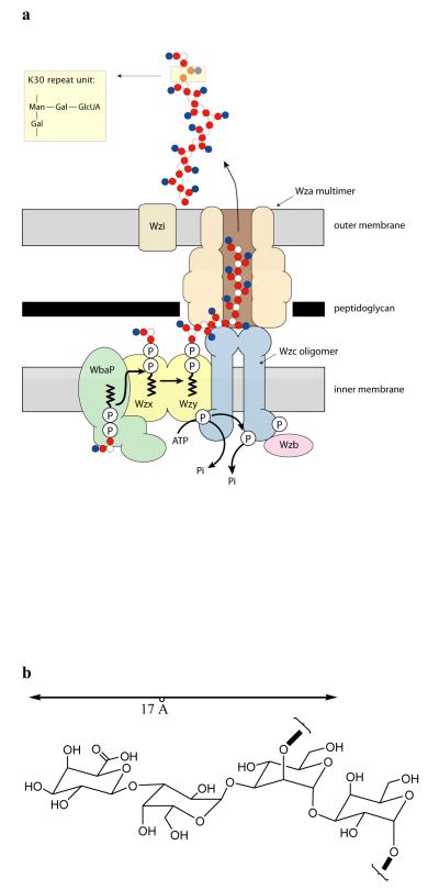
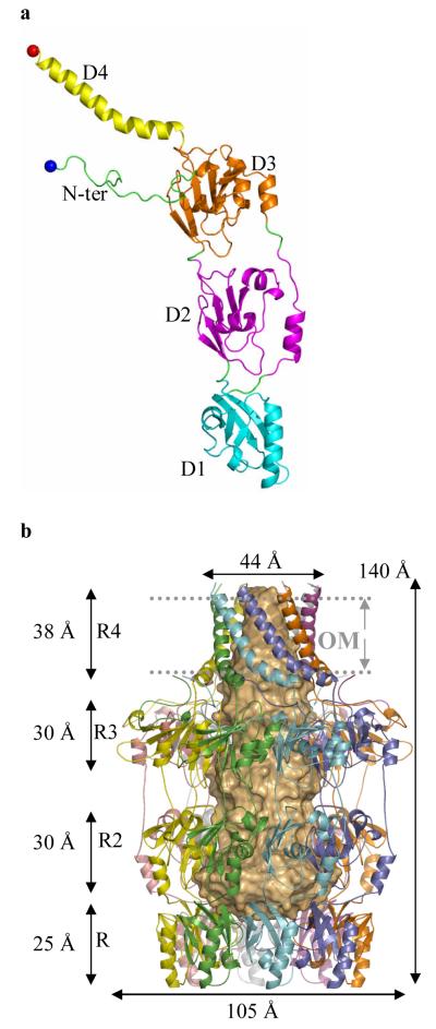
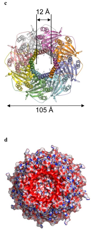
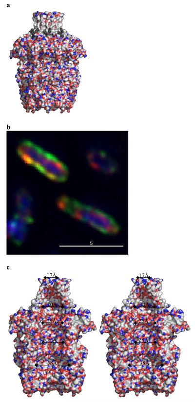
Many types of bacteria produce extracellular polysaccharides (EPSs). Some are secreted polymers and show only limited association with the cell surface, whereas others are firmly attached to the cell surface and form a discrete structural layer, the capsule, which envelopes the cell and allows the bacteria to evade or counteract the host immune system. EPSs have critical roles in bacterial colonization of surfaces, such as epithelia and medical implants; in addition some EPSs have important industrial and biomedical applications in their own right. Here we describe the 2.26 A resolution structure of the 340 kDa octamer of Wza, an integral outer membrane lipoprotein, which is essential for group 1 capsule export in Escherichia coli. The transmembrane region is a novel alpha-helical barrel. The bulk of the Wza structure is located in the periplasm and comprises three novel domains forming a large central cavity. Wza is open to the extracellular environment but closed to the periplasm. We propose a route and mechanism for translocation of the capsular polysaccharide. This work may provide insight into the export of other large polar molecules such as DNA and proteins.


Figures

Figure 1
Figure 1. Group 1 capsular polysaccharide export in Gram-negative bacteria
a, Model and proposed activities of a hypothetical biosynthetic complex carrying out coordinated synthesis and export of serotype K30 group 1 capsule in E. coli. Individual tetrasaccharide repeat units of the polymer are assembled by a series of enzymes including integral or peripheral membrane proteins on a lipid (undecaprenol diphosphate; und-PP) acceptor, using sugar nucleotide precursors available in the cytoplasm. The und-PP-linked repeat units are “flipped” across the inner membrane by a process involving an integral membrane protein (Wzx). Polymerization occurs at the periplasmic face and is dependent on another integral membrane protein, the putative polymerase, Wzy. High-level Wzy-dependent polymerization requires the activity of the tetrameric Wzc protein. To be active in capsule assembly, Wzc must undergo autophosphorylation. Dephosphorylation of Wzc by the Wzb phosphatase is also crucial for capsule synthesis, suggesting a need for cycling of the phosphorylation state of Wzc. Export of polymer to the surface requires the outer membrane Wza octameric complex,,. Biochemical and genetic data indicate that the Wza and Wzc proteins interact to form a complex that potentially spans the periplasm. b, A simple molecular model of the carbohydrate polymer, assuming the most extended conformation of the sugar, the maximum width that the channel has to accommodate at any point is approximately 17 Å.
Figure 2
Figure 2. The structure of Wza
a, The monomer of Wza can be decomposed into four domains; these are labeled as D1 - 4 and colored differently. Cys 21 (fatty acid modified N-terminus in mature Wza) is shown as a blue ball and Arg 376 (the last ordered residue) as a red ball. A stereo figure is shown in Supplementary Fig. S1a. b, The Wza octamer is shown in ribbon format. In this view the large central cavity is highlighted by space-filling light orange shape. The octamer is described as an amphora made of four rings (labeled R1-4). Ring 1 is formed by 8 copies of domain 1, ring 2 by 8 copies of domain 2 and so on. The helical barrel (R4) forms the “neck” of the structure and ring 1 (R1) the “base”. The predicted position of the outer membrane (OM) is marked and is found at the neck of the structure. The C-terminus of protein is predicted to be exposed on the cell surface and rings 1,2 and 3 located inside the periplasm. c, A view of the octamer such that one looks down into the central cavity from outside the cell through the helical barrel. The separation of loops which close the cavity in ring 1 are marked on the diagram. d, Wza is shown as a space filling model colored according to electrostatic charge and is rotated 180° from the view in Fig. 2c, such that one is now looking towards the cavity from periplasm. The concave surface of ring 1 can be seen and is closed by the loop at Tyr 110. There is a band of negative charge on the base of the structure. This may provide the association interface for protein-protein interactions with Wzc.
Figure 3
Figure 3. The central cavity of Wza
a, Wza is shown as a surface with polar atoms colored (colors are Fig. 2c.). Wza is oriented as the Fig. 2b. The lipid molecules are shown as black spheres and are located near the top of the structure. There are no gaps in the through the walls of the structure. The helical barrel is clearly non-polar and a band of tryptophan residues is exposed at the base of helical barrel (Fig S4b). b Green fluorescent anti-PK antibody binds to the surface of the cells which express Wza with a PK tag added to the C-terminus (Wza-PK). The periplasm is located by a red anti- alkaline phosphatase antibody and the nucleus is stained blue by DAPI. This confirms the C-terminus of Wza-PK is exposed on the surface of cells and thus the orientation of Wza is as shown in Fig. 2b. c, A stereo diagram of the internal cavity. The surface is colored according to polarity, (oxygen red, nitrogen blue and carbon, selenium, sulfur white) and reveals the interior of Wza is polar. The cavity is open at top through the helical barrel.

References

    1. Roberts IS. The biochemistry and genetics of capsular polysaccharide production in bacteria. Annu. Rev. Microbiol. 1996;50:285–315. - PubMed
    1. Donlan RM, Costerton JW. Biofilms: Survival mechanisms of clinically relevant microorganisms. Clin. Microbiol. Rev. 2002;15:167–193. - PMC - PubMed
    1. Hall-Stoodley L, Costerton JW, Stoodley P. Bacterial biofilms: From the natural environment to infectious diseases. Nat. Rev. Microbiol. 2004;2:95–108. - PubMed
    1. Fux CA, Costerton JW, Stewart PS, Stoodley P. Survival strategies of infectious biofilms. Trends Microbiol. 2005;13:34–40. - PubMed
    1. Sutherland IW. Novel and established applications of microbial polysaccharides. Trends Biotechnol. 1998;16:41–46. - PubMed

Publication types

MeSH terms

Substances

Associated data