. 2007 Mar 1;402(2):311-9.
doi: 10.1042/BJ20060861.

Ubc9 interacts with Lu/BCAM adhesion glycoproteins and regulates their stability at the membrane of polarized MDCK cells

Affiliations

Ubc9 interacts with Lu/BCAM adhesion glycoproteins and regulates their stability at the membrane of polarized MDCK cells

Emmanuel Collec et al. Biochem J. .

Abstract

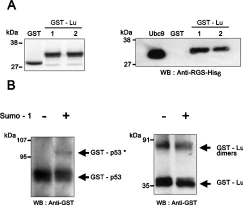
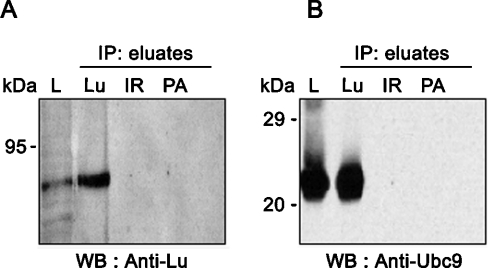
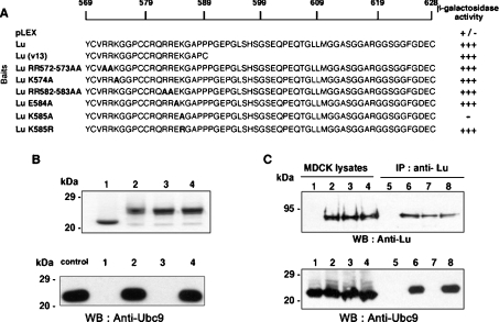
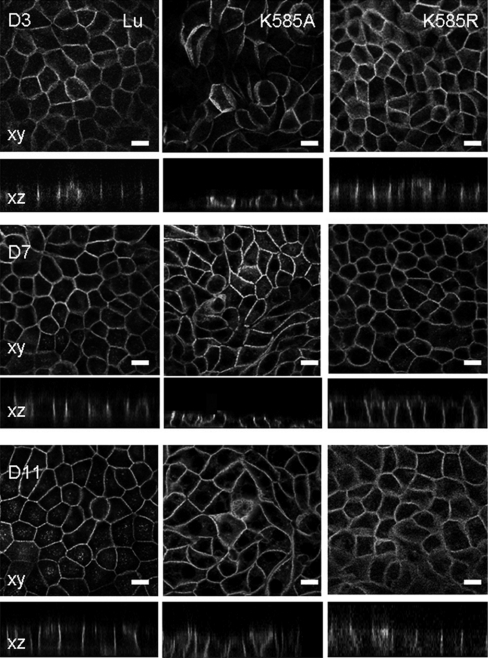
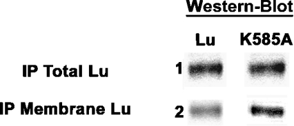
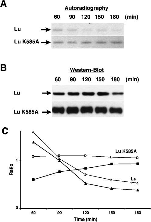
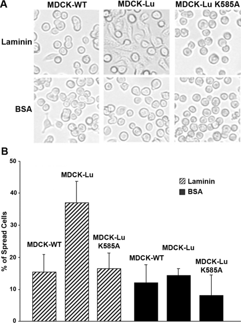
Lu (Lutheran) blood group and BCAM (basal cell adhesion molecule) antigens both reside on two gp (glycoprotein) isoforms, Lu and Lu(v13), that differ by the size of their cytoplasmic tail. They are receptors of laminin-10/11 and are expressed in RBCs (red blood cells), epithelial cells of multiple tissues and vascular endothelial cells. To gain more insights into the biological function of Lu/BCAM gps, we looked for potential partners of their cytoplasmic tail. We isolated Ubc9 (ubiquitin-conjugating enzyme 9) protein by screening a human kidney library using the yeast two-hybrid system. Lu/Ubc9 interaction was validated by GST (glutathione S-transferase) pull-down and co-immunoprecipitation experiments. Endogenous Ubc9 formed a complex with endogenous or recombinant Lu gp in A498 and MDCK (Madin-Darby canine kidney) epithelial cells respectively. Replacement of Lys(585) by alanine in the Lu gp abolished in vitro and ex vivo interactions of Lu gp with Ubc9 protein. Lu K585A mutant transfected in MDCK cells exhibited a normal basolateral membrane expression but was overexpressed at the surface of polarized MDCK cells as compared with wild-type Lu. Pulse-chase experiments showed extended half-life of Lu K585A gp at the plasma membrane, suggesting an impaired endocytosis of this mutant leading to protein accumulation at the membrane. Furthermore, we showed that the ability of MDCK-Lu K585A cells to spread on immobilized laminin was dramatically decreased. Our results support a physiological role for the direct interaction between Lu gp and Ubc9 protein and reveal a role for this enzyme in regulating the stability of Lu gp at the cell membrane.


Figures

Figure 1
Figure 1. In vitro interaction between Lu and Ubc9 studied by GST pull-down assays
(A) Left panel: Coomassie Blue staining showing that equivalent amounts of GST and GST–Lu were used for each analysis and that no degradation of GST proteins occurred during the assay. Right panel: His6-tagged Ubc9 protein was incubated with GST or GST–Lu in pull-down assays and the eluted proteins were analysed by Western blot (WB) using anti-RGS–His6 antibody. Two successive elutions were performed for GST–Lu interaction (lanes 1 and 2). An aliquot of the purified His6-tagged Ubc9 protein was loaded on to the gel as control (Ubc9 lane). (B) In vitro sumoylation assay with GST–Lu. GST–Lu was incubated with human Aos1/Uba2 (E1), UBC9 (E2) and with or without human SUMO-1. GST–p53 was used as positive control. GST proteins were detected by Western blot using a sheep anti-GST antibody. Sumoylated form of GST–p53* was detected at 96 kDa. No sumoylated form of GST–Lu was detected at the expected molecular mass (53 kDa). Dimers of GST–Lu were detected at 70 kDa.
Figure 2
Figure 2. Ex vivo interaction of Lu and Ubc9 by co-IP
A498 cells (107) were lysed and extracts were subjected to IP with monoclonal LM342 anti-Lub (Lu), irrelevant antibody (IR) and Protein A–Sepharose (PA). Presence of Lu gps (A) and Ubc9 (B) were studied by Western blot (WB) in lysates (L) and in IP eluates using a rabbit anti-Lu (602) and a sheep anti-human Ubc9 antibodies respectively.
Figure 3
Figure 3. Mapping of the Ubc9-binding site in the cytoplasmic tail of Lu gp
(A) Yeast two-hybrid experiments. Yeast was co-transformed with pACT2-Ubc9 and pLEX (DNA-binding domain alone), pLEX-Lu, -Lu(v13), -Lu RR572–573AA, -Lu K574A, -Lu RR582–583AA, -Lu E584A, -Lu K585A, or -Lu K585R. β-Galactosidase activity was determined on filters for each construct. Mutated amino acids are in bold type. (B) GST pull-down assays. Upper panel: Coomassie Blue staining was used to show that equivalent amounts of GST (1), GST–Lu (2), GST–Lu K585A (3) and GST–Lu K585R (4) were used. Lower panel: His6-tagged Ubc9 protein was incubated with GST (1), GST–Lu (2), GST–Lu K585A (3) or GST–Lu K585R (4) in pull-down assays. Proteins were eluted and analysed by Western blot (WB) using a sheep anti-Ubc9 antibody. An aliquot of the purified His6-tagged Ubc9 protein was loaded on to the gel as control (‘control’ lane). (C) IP of Lu gp in MDCK cells. Wt (1 and 5) and transfected MDCK clones expressing Lu gp (2 and 6) or mutants Lu K585A (3 and 7) or Lu K585R (4 and 8) were lysed and extracts were subjected to IP with monoclonal LM342 anti-Lub antibody. Presence of Lu gp (upper panel) and Ubc9 (lower panel) were tested by Western blot (WB) in lysates (1–4) and in IP eluates (5–8) using a rabbit polyclonal anti-Lu (602) and a sheep Ubc9 antibodies respectively.
Figure 4
Figure 4. Immunofluorescence studies of the Lu mutants in MDCK cells
Stably transfected MDCK-Lu gp, -Lu K585A and -Lu K585R cells were grown on filters from day 3 (D3) up to day 11 (D11). Confocal imaging shows fluorescence staining of Lu gp in xy (en face view) and xz (side views) sections. Scale bars, 10 μM.
Figure 5
Figure 5. Expression level of Lu and Lu K585A mutant at the membrane of polarized MDCK cells grown on Transwell filters
Western blot using rabbit polyclonal anti-Lu antibody (602) showing the total amounts of Lu and Lu K585A gps after IP using mouse anti-Lu antibody (F241) compared with the biotinylated Lu and Lu K585A gps expressed at the membrane.
Figure 6
Figure 6. The expression level and the turnover of Lu and Lu K585A mutant at the membrane of polarized MDCK cells
MDCK cells expressing Lu and Lu K585A were grown on Transwell filters. (A) Newly delivered Lu and Lu K585A gps at the cell surface as determined by autoradiography of biotinylated Lu gps. (B) Western blot showing total amounts of Lu and Lu K585A gps expressed at the membrane at each time of chase, using rabbit polyclonal anti-Lu antibody (602). (C) The curves show the ratio of ‘radiolabelled biotinylated Lu/total biotinylated Lu’ at the membrane reflecting the turnover of newly delivered Lu and Lu K585A gps at the cell surface as a function of time. The curves represent mean values obtained with cellular pools (Lu: △, n=2; Lu K585A: □, n=2) and clones (Lu: ▲, n=4; Lu K585A: ■, n=4).
Figure 7
Figure 7. Morphological cell adhesion assay of MDCK-WT, -Lu and -Lu K585A cells to laminin-10/11
MDCK-WT, -Lu or -Lu K585A cells (105) were incubated at 37 °C for 3 h in wells coated with 2 μg/ml of laminin-10/11 or 1% BSA. (A) Photos showing cell morphology of each cell line. (B) The histogram presents the percentage of spread cells after 3 h of adhesion on laminin-10/11 or BSA. Results represent the means for four experiments.

Similar articles

Cited by

References

    1. Cartron J. P., Colin Y. Structural and functional diversity of blood group antigens. Transfus. Clin. Biol. 2001;8:163–199. - PubMed
    1. Daniels G. Terminology for red cell antigens – 1999 update. Immunohematology. 1999;15:95–99. - PubMed
    1. El Nemer W., Rahuel C., Colin Y., Gane P., Cartron J. P., Le Van Kim C. Organization of the human LU gene and molecular basis of the Lu(a)/Lu(b) blood group polymorphism. Blood. 1997;89:4608–4616. - PubMed
    1. Rahuel C., Le Van Kim C., Mattei M. G., Cartron J. P., Colin Y. A unique gene encodes spliceoforms of the B-cell adhesion molecule cell surface glycoprotein of epithelial cancer and of the Lutheran blood group glycoprotein. Blood. 1996;88:1865–1872. - PubMed
    1. El Nemer W., Gane P., Colin Y., Bony V., Rahuel C., Galacteros F., Cartron J. P., Le Van Kim C. The Lutheran blood group glycoproteins, the erythroid receptors for laminin, are adhesion molecules. J. Biol. Chem. 1998;273:16686–16693. - PubMed

Publication types

MeSH terms

Substances