. 2006 Nov;25(5):795-806.
doi: 10.1016/j.immuni.2006.09.012.

ERAAP synergizes with MHC class I molecules to make the final cut in the antigenic peptide precursors in the endoplasmic reticulum

Affiliations

ERAAP synergizes with MHC class I molecules to make the final cut in the antigenic peptide precursors in the endoplasmic reticulum

Takayuki Kanaseki et al. Immunity. 2006 Nov.

Abstract

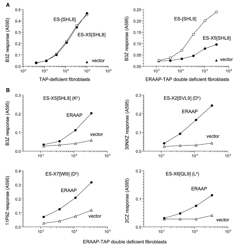
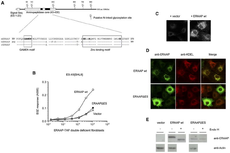
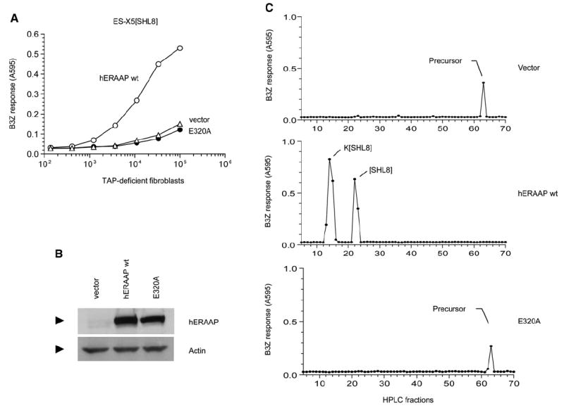
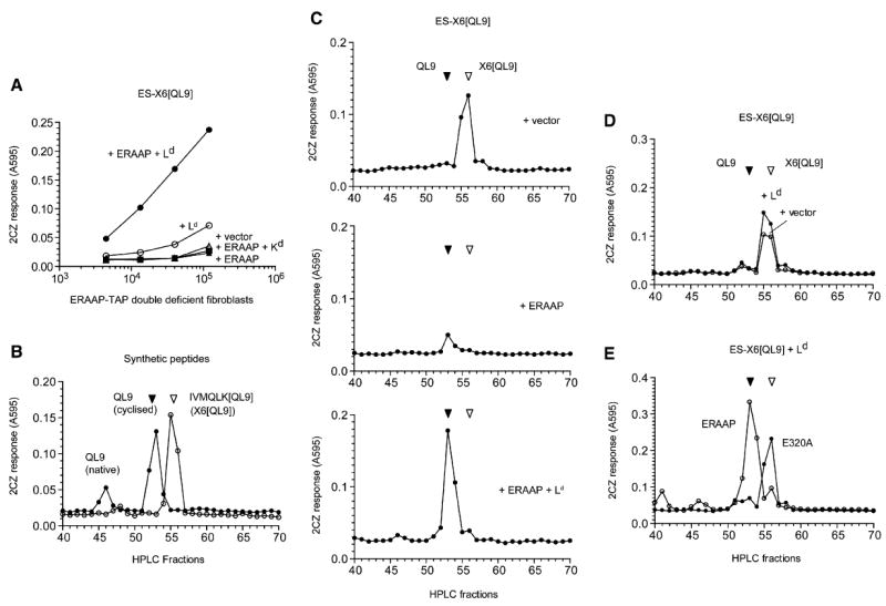
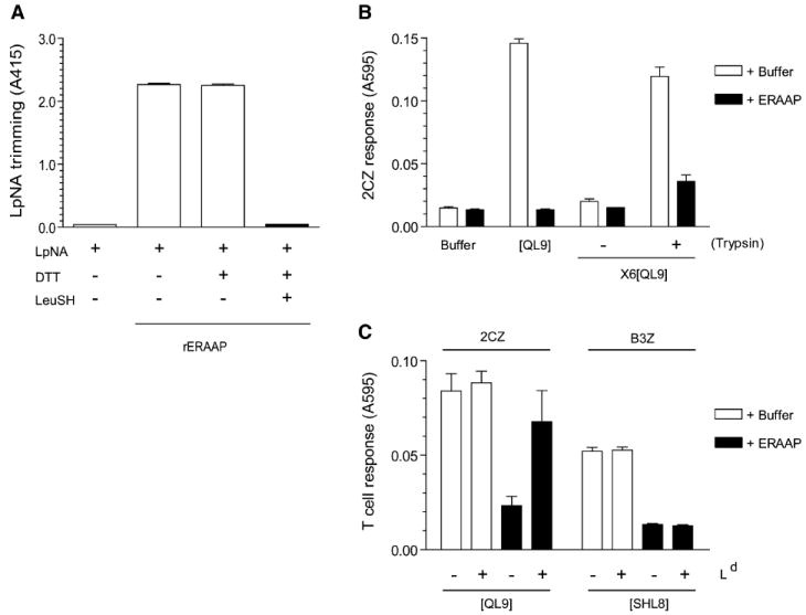
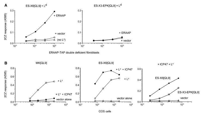
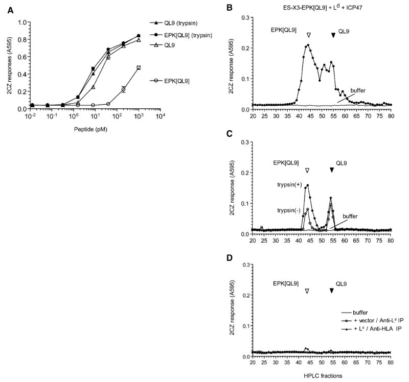
The major histocompatibility complex class I molecules display peptides (pMHC I) on the cell surface for immune surveillance by CD8(+) T cells. These peptides are generated by proteolysis of intracellular polypeptides by the proteasome in the cytoplasm and then in the endoplasmic reticulum (ER) by the ER aminopeptidase associated with antigen processing (ERAAP). To define the unknown mechanism of ERAAP function in vivo, we analyzed naturally processed peptides in cells with or without appropriate MHC I and ERAAP. In the absence of MHC I, ERAAP degraded the antigenic precursors in the ER. However, MHC I molecules could bind proteolytic intermediates and were essential for generation of the final peptide by ERAAP. Thus, ERAAP synergizes with MHC I to generate the final pMHC I repertoire.


Conflict of interest statement

The authors declare that they have no conflicts of interest.

Figures

Figure 1
Figure 1. ERAAP Is Essential for Generating pMHC I on the Cell Surface from ER-Targeted N-Terminally Extended Precursors
(A) The DNA constructs encoding the vector alone or the SHL8 (SIINFEHL) antigenic peptide downstream of an ER-targeting signal sequence (ES) were transiently transfected into fibroblasts derived from either TAP-deficient or ERAAP-TAP double-deficient mice. The constructs included either no (ES-[SHL8]) or five (ES-X5[SHL8], X5 = AIVMK) additional amino acids between the signal peptidase cleavage site of the ES-signal sequence and the SHL8 peptide. 2 days later, indicated number of transfected cells were incubated overnight with the lacZ-inducible, SHL8-Kb-specific B3Z T cell hybridoma. The lacZ activity was measured by the conversion of the substrate chlorophenolred-β-D-galacto-pyrannoside into chlorophenol red, which absorbs light at 595 nm. (B) ER-targeted, N-terminally extended versions of the antigenic peptides SHL8, SVL9 (SSVVGVWYL), WI9 (WMHHNMDLI), or QL9 (QLSPFPFDL) were cotransfected with either vector alone or mouse ERAAP into ERAAP-TAP double-deficient fibroblasts. The cDNA encoding Ld was also included with the ES-X6[QL9] construct. 2 days later, varying number of transfected cells were used as APCs for the indicated T cells, and their lacZ response was measured as in (A). The MHC molecule presenting the peptides is shown in parentheses. Data are representative of at least three independent experiments.
Figure 2
Figure 2. The Generation of pMHC I from N-Terminally Extended Precursors Requires ERAAP Expression in the ER Compartment
(A) Schematic of mouse ERAAP containing the putative ER translocation signal (ES), the aminopeptidase core, and potential N-linked glycosylation sites. The sequence alignment of the aminopeptidase core with the human and rat orthologs is shown with the conserved GAMEN and zinc binding motifs (boxed). (B) Deletion of the ES sequence eliminates ERAAP function. The wild-type ERAAP (ERAAP WT), its ES deletion mutant (ERAAPΔES), or vector alone were cotransfected with the ER-targeted antigenic precursor ES-X9[SHL8]. Surface expression of SHL8-Kb complex was measured with B3Z T cells. (C) Polyclonal rabbit anti-mouse ERAAP antibodies specifically stain ERAAP-transfected COS cells. The COS cells were transfected with either vector or ERAAP cDNA and stained with the primary and secondary antibodies 2 days later as described in the Experimental Procedures. (D) Deletion of ES signal alters the intracellular location of ERAAP from the ER to the cytoplasm. COS cells transfected with either ERAAP WT or ERAAPΔES were stained with the ERAAP (green) or the ER marker KDEL (red) antibody. The merged image shows an overlay of green and red stains as yellow. (E) ERAAP without the ES signal remains unglycosylated. The extracts of COS cells transfected with ERAAP WT or ERAAPΔES were either untreated or treated with Endo H. The samples were separated by SDS-PAGE, transferred to the nitrocellulose membranes, and immunoblotted with the ERAAP antibody. Data are representative of at least two independent experiments.
Figure 3
Figure 3. The Generation of pMHC I from N-Terminally Extended Precursors in the ER Requires Enzymatically Active ERAAP
(A) The cDNAs encoding either vector alone, human ERAAP WT, or its single amino acid mutant in the GAMEN motif (E320A) were cotransfected into ERAAP-TAP double-deficient fibroblasts together with the ES-X5[SHL8] construct. After 2 days, expression of SHL8-Kb complex was measured with B3Z T cells. (B) The hERAAP WT and its E320A mutant are expressed at comparable amounts. The amounts of hERAAP WT, its E320A mutant, or vector alone as a negative control were determined by immunoblotting transfected COS cell lysates with the hERAAP antibody. (C) The conversion of the N-terminally extended precursor to the final SHL8 or K[SHL8] peptides presented by Kb or Db MHC occurs only in the presence of hERAAP WT. The peptide extracts from ERAAP-TAP double-deficient fibroblasts transfected with ES-X5[SHL8] and either vector alone, hERAAP WT, or its E320A mutant were fractionated by RP-HPLC. The SHL8-containing, N-terminally extended intermediates were detected after trypsin treatment of each fraction and incubating them with B3Z T cells in the presence of the appropriate APC. The peaks were identified, where known, by comparison with synthetic peptides run under identical conditions. Data are representative of at least two independent experiments.
Figure 4
Figure 4. The Fate of N-Terminally Extended X6[QL9] and the Final QL9 Peptide Is Determined by ERAAP and the Ld MHC Molecule
(A) Generation of the QL9-Ld complex in ERAAP-TAP double-deficient fibroblasts from the ER-targeted ES-X6[QL9] precursor requires ERAAP and the Ld MHC. The ERAAP-TAP double-deficient fibroblasts were cotransfected with the ES-X6[QL9] precursor and other cDNAs shown. Expression of the QL9-Ld complex was detected with lacZ-inducible 2CZ T cell hybridoma as in Figure 1. (B) Synthetic IVMQLK[QL9] and the QL9 peptides can be resolved by HPLC and detected by 2CZ T cells. 10 fmoles of indicated peptides were injected into the HPLC. Each fraction was treated with trypsin and assayed with 2CZ T cells and Ld-L cells as APCs. (C) ERAAP generates the final QL9 peptide from the X6[QL9] precursor in the presence of Ld but degrades the precursor peptide in the absence of Ld. The ERAAP-TAP double-deficient fibroblasts were cotransfected with the ES-X6[QL9] precursor construct and either vector alone, ERAAP alone, or ERAAP plus Ld. The peptides were extracted from the transfected cells and fractionated by HPLC. Each fraction was treated with trypsin and used to stimulate 2CZ T cells in the presence of Ld-L cells as APCs. (D) The absence of ERAAP cannot be compensated by other proteases. The ERAAP-TAP double-deficient fibroblasts were transfected with the ES-X6[QL9] construct with either vector alone or Ld, and cell extracts were analyzed for 2CZ activation after HPLC fractionation and trypsin treatment of each fraction. (E) Generation of the final QL9 peptide from its X6[QL9] precursor in the presence of Ld requires enzymatically active ERAAP. The ERAAP-TAP double-deficient fibroblasts were cotransfected with the ES-X6[QL9] precursor, Ld, and either hERAAP WT or its enzymatically inactive mutant E320A. The cell extracts were analyzed for 2CZ activation after HPLC fractionation and trypsin treatment. Data are representative of at least three (C) or two independent experiments.
Figure 5
Figure 5. Recombinant ERAAP Trims the Final QL9 as well as Its X6[QL9] Precursor In Vitro, and Ld MHC Can Protect QL9 Peptide
(A) Purified recombinant ERAAP (rERAAP) is active in trimming the model LpNA substrate, and leucinethiol inhibits this activity. The rERAAP purified from baculovirus-infected insect cells was incubated with Leucine pnitroanilide (LpNA) with or without leucinethiol (LeuSH) and DTT, which is required to keep LeuSH in its reduced and active form. The LpNA hydrolysis was measured as light absorbance at 415 nm. (B) The indicated synthetic peptides were incubated with either buffer alone or with rERAAP. The peptides were assayed by their ability to stimulate 2CZ T cells in the presence of Ld-L cells as APCs. To detect the generation of QL9 peptide from its N-terminally extended X6[QL9] precursor, the peptides were either assayed as such or after trypsin treatment. (C) The Ld MHC protects QL9 but not SHL8 peptide from degradation by rERAAP. Synthetic QL9 or SHL8 peptides were incubated with buffer or rERAAP in the presence or absence of recombinant Ld-Ig fusion protein. The QL9 and SHL8 peptides were assayed with 2CZ or B3Z T cells and Ld or Kb-L cells as APCs, respectively. The graph shows the mean values with SD from three independent experiments (A–C).
Figure 6
Figure 6. Model for Detecting MHC I-Bound Antigentic Precursors
(A) The presence of a proline residue among the N-terminal flanking residues inhibits ERAAP’s ability to generate the final QL9-Ld complex. The ERAAP-TAP double-deficient fibroblasts or COS cells were cotransfected with ER-targeted, N-terminally extended QL9 precursors without (ES-X6[QL9]) or with (ES-X3-EPK[QL9]), a proline residue (P), the Ld MHC, and either ERAAP or vector alone. The presence of the QL9-Ld complex on the cell surface was measured with 2CZ T cells. (B) The TAP inhibitor ICP47 blocks presentation of cytoplasmic but not ER-targeted QL9 precursors in COS cells. Simian COS cells were cotransfected with constructs encoding cytoplasmic (MK[QL9]) or ER-targeted (ES-X6[QL9], ES-X3-EPK[QL9]) precursors, and the indicated cDNAs encoding Ld, ICP47, or vector alone. The presence of the QL9-Ld complex on the cell surface was measured with 2CZ T cells. Data are representative of at least two independent experiments. The similar results were confirmed by using either ES-X3-LPK[QL9] or -QPK[QL9] instead of -EPK[QL9] (data not shown).
Figure 7
Figure 7. The N-Terminally Extended EPK[QL9] Proteolytic Intermediate Is Bound by the Ld MHC Molecules
(A) Trypsin treatment of N-terminally extended QL9 precursor EPK[QL9] enhances its antigenicity. Varying concentrations of synthetic QL9 and its N-terminally extended analog EPK[QL9] peptide were tested for their ability to stimulate 2CZ T cells with Ld-L cells as APC. The peptides were tested as such or after preincubation with trypsin. (B) The final antigenic QL9 peptide, its N-terminally extended precursors and intermediates present in the total peptide extract of COS cells transfected with ES-X3-EPK[QL9], Ld, and ICP47. The extract was fractionated by HPLC and antigenic peptides detected in the fractions by their ability to stimulate 2CZ response after trypsin treatment. HPLC fractions collected after injection of buffer alone were tested in parallel to rule out carryover between samples. (C) The Ld MHC was immunoprecipitated from detergent lysate of COS cells transfected with the ES-X3-EPK[QL9] construct, Ld, and ICP47 cDNAs. The peptides contained in the immunoprecipitated material were fractionated by HPLC and assayed with 2CZ T cells with (closed circle) or without (open circle) trypsin treatment. (D) As controls, the same cell lysate was immunoprecipitated with the pan HLA (W6/32) antibody (triangle), or the lysate from cells without Ld transfection was immunoprecipitated with the Ld antibody (circle). The peptide content of the immunoprecipitated material was analyzed as above. Data are representative of at least three independent experiments.

Similar articles

Cited by

References

    1. Brouwenstijn N, Serwold T, Shastri N. MHC class I molecules can direct proteolytic cleavage of antigenic precursors in the endoplasmic reticulum. Immunity. 2001;15:95–104. - PubMed
    1. Cascio P, Hilton C, Kisselev AF, Rock KL, Goldberg AL. 26S proteasomes and immunoproteasomes produce mainly N-extended versions of an antigenic peptide. EMBO J. 2001;20:2357–2366. - PMC - PubMed
    1. Chang SC, Momburg F, Bhutani N, Goldberg AL. The ER aminopeptidase, ERAP1, trims precursors to lengths of MHC class I peptides by a “molecular ruler” mechanism. Proc Natl Acad Sci USA. 2005;102:17107–17112. - PMC - PubMed
    1. Corr M, Boyd LF, Frankel SR, Kozlowski S, Padlan EA, Margulies DH. Endogenous peptides of a soluble major histocompatibility complex class I molecule, H-2Lds: sequence motif, quantitative binding, and molecular modeling of the complex. J Exp Med. 1992;176:1681–1692. - PMC - PubMed
    1. Elliott T, Townsend A, Cerundolo V. Antigen presentation: naturally processed peptides. Nature. 1990;348:195–197. - PubMed

Publication types

MeSH terms

Substances