Comparative Study
. 2006 Nov 9;52(3):425-36.
doi: 10.1016/j.neuron.2006.08.028.

Refined spatial manipulation of neuronal function by combinatorial restriction of transgene expression

Affiliations
Comparative Study

Refined spatial manipulation of neuronal function by combinatorial restriction of transgene expression

Haojiang Luan et al. Neuron. .

Abstract

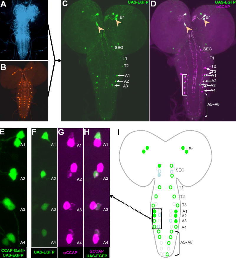
Selective genetic manipulation of neuronal function in vivo requires techniques for targeting gene expression to specific cells. Existing systems accomplish this using the promoters of endogenous genes to drive expression of transgenes directly in cells of interest or, in "binary" systems, to drive expression of a transcription factor or recombinase that subsequently activates the expression of other transgenes. All such techniques are constrained by the limited specificity of the available promoters. We introduce here a combinatorial system in which the DNA-binding (DBD) and transcription-activation (AD) domains of a transcription factor are independently targeted using two different promoters. The domains heterodimerize to become transcriptionally competent and thus drive transgene expression only at the intersection of the expression patterns of the two promoters. We use this system to dissect a neuronal network in Drosophila by selectively targeting expression of the cell death gene reaper to subsets of neurons within the network.


Figures

Figure 1
Figure 1. The ternary Split Gal4 System improves upon existing binary expression systems in restricting transgene expression
(A) Schematic depiction of the Gal4-UAS system of Drosophila, a classic binary system for targeting transgene expression in vivo. The first essential component of this system, shown at left, is a transgene containing the yeast transcription factor Gal4 downstream of a promoter/enhancer (P). P drives expression of Gal4 in flies bearing this transgene in a cell-type specific manner (shown as a black region within the CNS). When flies bearing Gal4 are crossed to flies bearing the second component of the system, a transgene of interest placed downstream of the Gal4 DNA recognition site or UAS, this transgene is also expressed in the same cell-type specific manner (right). (B) The Split Gal4 system exploits the fact that the two functional domains of Gal4, the DNA binding (DBD) and transcription activation (AD) domains, are separable. In the Split Gal4 system each domain is fused to a heterodimerizing leucine zipper (Zip+ or Zip) to insure that the two domains associate when expressed in the same cell and reconstitute transcriptional activity. Starting at left, the schematic shows that the Gal4DBD and AD constructs can be independently targeted using different promoters/enhancers (P1 and P2). When these constructs (“hemidrivers”) are brought together in crosses to flies bearing a UAS-transgene, the transgene is expressed in progeny only at the intersection of the expression patterns of P1 and P2 (designated by the intersection sign, ∩ ), where transcriptional activity bottom right. The Gal4DBD must be used if UAS-transgenes are to be transcribed, but any transcription activation domain can be used, as long as it is fused to the leucine zipper fragment complementary to that fused to Gal4DBD.
Figure 2
Figure 2. Optimized Split Gal4 constructs efficiently drive UAS-reporter gene expression in vitro and in vivo in the Drosophila nervous system
(A) Schematic representation of the three optimized Split Gal4 constructs: (top) the DNA-binding domain of the Gal4 molecule fused at the N-terminus to the Zip leucine zipper (see Experimental Procedures), (middle) the major transcription activation domain of Gal4 fused at the C-terminus to the Zip+ leucine zipper (Gal4AD), and (bottom) a similar AD construct containing the C-terminal acidic domain of the VP16 transcription factor. The sawtooth line represents a flexible linker of 10 consecutive glycine residues. NLS, SV40 nuclear localization signal. The Gal4DBD contains an endogenous N-terminal NLS (not depicted). (B) Relative transcriptional activities of the Split Gal4 constructs when expressed in cultured cells, alone and in combination. Transcriptional activity was measured indirectly as the ß-galactosidase activity in lysates of Drosophila SL-2 cells co-transfected with plasmids containing the indicated Split Gal4 constructs and a UAS-LacZ gene. ß-galactosidase activity was normalized to the total protein and is expressed as the percentage of the activity measured in cells transfected with intact Gal4. Bars show average activity in three transfections. (C–H) Fluorescence photomicrographs of Drosophila 3rd instar larval CNS from animals expressing (C) Gal4DBD alone, (D) VP16AD alone, (E) Gal4DBD alone (F) Gal4DBD and Gal4AD, (G) intact Gal4, or (H) Gal4DBD and VP16AD. Expression was driven by the pan-neuronal elav promoter. Fluorescence shows expression of one or two copies of a UAS-EGFP reporter transgene, as indicated. (E) Long exposure image (6-fold longer than for the other panels) reveals weak expression of the reporter driven by the Gal4DBD construct alone. Here and elsewhere, hemidrivers are denoted by the promoter used to drive the (superscripted) Split Gal4 construct, with Gal4DBD abbreviated to DBD, and ∩ denotes the combination of hemidrivers expressed in a cross.
Figure 3
Figure 3. The Split Gal4 system restricts UAS-transgene expression to the intersection of the expression patterns of two promoters
Comparison of UAS-EGFP expression in the CNS of 3rd instar larvae driven by (A–B) CCAP-Gal4, or (C–F) combined Split Gal4 hemidrivers made with the pan-neuronal elav promoter and the promoter for the CCAP gene. (A) UAS-EGFP expression pattern of CCAP-Gal4 driver. This pattern consists of 48 neurons that express the neuropeptide CCAP. (B) Consensus expression pattern derived from analysis of 6 preparations like the one shown in (A), as described in Experimental Procedures. (C–D) UAS-EGFP expression pattern driven by elavGal4DBD ∩ CCAPGal4AD and the corresponding consensus pattern (n=6). (E–F) UAS-EGFP expression pattern driven by CCAPGal4DBD ∩ elavVP16AD and the corresponding consensus pattern (n=5). Consensus pattern symbols: circles, the canonical CCAP–expressing neurons, showing their anatomical positions in the CNS (Br, brain; SEG, subesophageal ganglion; T1-3, the three thoracic ganglia; A1–A8, the eight abdominal ganglia); green circles, neurons that expressed EGFP in greater than two-thirds of the preparations; gray circles (see A8 in D and F), neurons that expressed EGFP in less than two-thirds of preparations. Solid green circles: high EGFP expression; open green circles, low EGFP expression. A few “ectopically” labeled CCAP-immunonegative cells were observed for each driver or hemidriver combination. Their average number per preparation (± SD) was 3±2 (B), 4±2 (D) and 2±2 (F).
Figure 4
Figure 4. Genetic ablation of NCCAP neurons using the Split Gal4 system causes pupal lethality and wing expansion deficits that correlate with expression strength
(A) Cumulative percentage of progeny displaying the pupal lethal (black bars) and unexpanded wing (gray bars) phenotypes, following ablation of neurons within the CCAP expression pattern by targeted expression of one or two copies of the cell death gene reaper using the indicated driver or combination of hemidrivers. If these phenotypes were not present, no bar is shown. *, embryonic or early larval lethality. (Inset) Adult wing phenotypes. (Left) Unexpanded wings of a newly emerged adult fly. (Right) Same fly after expanding its wings. Adult flies lacking the CCAP-expressing neurons permanently retain the unexpanded wing phenotype. (B–E) Confocal micrographs of CCAP-immunoreactive neurons in the nervous systems of pharate adult flies expressing UAS-reaper driven by the indicated drivers or hemidriver combinations. (B) CCAP-Gal4, (C) elavGal4DBD and CCAPGal4AD, (D) CCAPGal4DBD and elavVP16AD, and (E) CCAPGal4DBD alone. The CCAP-immunopositive neurons shown survived the developmental expression of reaper. The average number of surviving NCCAP neurons (± SD) in preparations like those shown in panels B, C, D, and E were: 6.6 ± 0.9 (n=5), 15.3 ± 1.5 (n=9), 0±0 (n=4), and 47.6 ± 0.5 (n=5), respectively. Abbreviations as in Fig. 3, except AG, fused abdominal ganglion of the pharate adult CNS. On average, the pharate adult AG has two fewer NCCAP neurons than are found in the larval abdominal ganglia. Also, the identity of NCCAP neurons in the pharate adult brain is unlikely to correspond to those found in the larval brain.
Figure 5
Figure 5. A subset of NCCAP neurons lies within the expression pattern of the choline acetyltransferase (Cha) promoter, as identified by Cha-Gal4>UAS-EGFP labeling
(A) Consensus labeling pattern showing the overlap of Cha-Gal4 expression (detected with UAS-EGFP) with NCCAP (detected by anti-CCAP immunolabeling; n=9). Circles, CCAP-immunopositive neurons. Green circles, EGFP labeled in greater than one-third of the preparations. Light blue circles, EGFP labeled in one-third or less of the preparations. Gray circles, neurons that were never EGFP-labeled. Filled circles, consistently high EGFP expression (see Experimental Procedures). Open circles, low EGFP expression. In paired NCCAP neurons in hemisegments T3–A4, one was typically strongly CCAP-immunopositive and the other was more weakly labeled. For convenience, we have shown the strongly expressing neuron as medial to the weakly expressing one. This does not necessarily represent their anatomical positions. (B–J) High-resolution confocal micrographs showing anti-CCAP immunoreactivity (B, E, H), EGFP expression (C, F, I), and their overlap (D, G, J). For each set of images, individual neurons are indicated by arrows and dotted outlines. Boxes in (A) show the anatomical location of the corresponding images. In hemisegments T3 and A1 to A4, only the more weakly CCAP immunoreactive neuron of the pair expresses EGFP.
Figure 6
Figure 6. Targeting the cholinergic subset of NCCAP, using complementary hemidrivers made with the Cha and CCAP promoters
The pattern of UAS-EGFP expression in the CNS of Drosophila 3rd instar larvae driven by the Split Gal4 hemidrivers ChaGal4DBD and CCAPGal4AD. (A–B) Expression patterns of the promoters used to make the ChaGal4DBD and CCAPGal4AD hemidriver lines. (A) Cha-Gal4>UAS-EGFP (blue pseudocolor). (B) CCAP-Gal4>UAS-EGFP (red pseudocolor). (C–D) Restricted pattern of UAS-EGFP expression within NCCAP in ChaGal4DBD ∩ CCAPGal4AD crosses. (C) A representative example of EGFP expression. (D) Both EGFP expression (green) and CCAP-immunostaining (magenta) in sample from (C). Anatomical abbreviations are as in Fig. 3. Consistently double-labeled neurons in the brain (arrowheads) and in hemisegments A1–A4 (box in D) are indicated. (E) UAS-EGFP driven by CCAP-Gal4 expresses in both strong and weak CCAP-immunoreactive cells of hemisegments A1–A4. (F–H) Higher magnification images of the boxed region in (D) showing restriction of EGFP expression to only the more weakly immunoreactive NCCAP neuron of the pair in hemisegments A1–A4. (F) UAS-EGFP expression. (G) CCAP-immunolabeling. (H) Merged image of (F) and (G). (I) Consensus pattern of UAS-EGFP expression driven by ChaGal4DBD ∩ CCAPGal4AD, derived from seven preparations. The average frequency and intensity of labeling for eachidentified neuron are designated as in Fig. 5. Boxed region, hemisegments A1–A4 shown in (F–H). Ectopically labeled neurons are not represented in the consensus pattern. On average 7±2 (n=7) non-CCAP-immunoreactive cells were observed per preparation. Some of these, such as the two in the brain, appear to correspond to cells also sometimes seen in CCAP-Gal4 labeling patterns.
Figure 7
Figure 7. Split Gal4 enhancer-trap lines can be used to target distinct subsets of neurons within NCCAP
Patterns of EGFP expression in the CNS of Drosophila 3rd instar larvae driven by the CCAPGal4DBD hemidriver combined with two different VP16AD enhancer trap lines. (A–C) Expression patterns of two VP16AD enhancer trap hemidrivers and CCAP-Gal4. Nervous system expression (blue pseudocolor) of (A) the ETVP16AD-N4 and (C) ETVP16AD-N6 enhancer-trap lines revealed by combining them with the elavGal4DBD hemidriver in the presence of a UAS-EGFP reporter. (B) CCAP-Gal4>UAS-EGFP. (D–G) Restricted patterns of UAS-EGFP expression generated by (D) CCAPGal4DBD ∩ ETVP16AD-N4, and (F) CCAPGal4DBD ∩ ETVP16AD-N6, and (E, G) their corresponding consensus patterns. Consensus patterns were derived from 4 (ETVP16AD-N4) and 6 ETVP16AD-N6) double-labeled preparations, respectively, with neurons represented as in Fig. 5.
Figure 8
Figure 8. Genetic ablation of neurons within NCCAP using the Split Gal4 system identifies anatomically and functionally distinct subsets of this network
(A–D) Pattern of NCCAP neuronal survival after ablation by UAS-reaper, driven by CCAPGal4DBD ∩ ETVP16AD-N4. (A) Confocal image showing surviving CCAP-immunopositive cells in the CNS of a pharate adult. (B) Pattern of bursicon immunoreactivity in the same double-labeled preparation. (C) Merged image of (A) and (B). The average number of surviving NCCAP neurons (± SD) from seven preparations was 30.3 ± 2.1. (D) Consensus pattern of surviving (red filled circles) and ablated (black open circles) NCCAP neurons. Note that ETVP16AD-N4 expression within NCCAP changes developmentally so that more neurons are killed than are seen in the larval CNS expression pattern (Fig. 7E). ET–N4, enhancer-trap line ETVP16AD-N4. (E–H) NCCAP neuronal survival after ablation by UAS-reaper, driven by CCAPGal4DBD ∩ ETVP16AD-N6, similar to (A–D). The average number of surviving NCCAP neurons (± SD) from eight preparations was 32.9 ± 5.6. ET–N6, enhancer-trap line ETVP16AD-N6. (I) The patterns of anti-CCAP (magenta) and anti-bursicon (green) labeling in the CNS of a control animal are shown for comparison. The pharate adult CNS typically has 48 NCCAP neurons, including 14 bursicon-immunopositive neurons in the AG and 1 or 2 pairs in the SEG. (J) Percentage of progeny with unexpanded wings in crosses with UAS-reaper driven by CCAPGal4DBD ∩ ETVP16AD-N4 and CCAPGal4DBD ∩ ETVP16AD-N6. Gray bar shows the frequency of the unexpanded wing phenotype. No bar denotes normally expanded wings. (K) A critical subset (red filled black circles) of neurons within NCCAP, some or all of which must be necessary for wing expansion. This subset includes all neurons ablated in crosses with ETVP16AD-N4 that were not also ablated in crosses with ETVP16AD-N6. Neurons ablated with ETVP16AD-N6 are clearly not necessary for wing expansion since flies from crosses with this hemidriver showed normal wing expansion. The critical set does not contain any of the bursicon-expressing neurons in the AG. Because the identities of the non-bursicon-expressing neurons in the abdominal ganglion are difficult to determine, it is unclear whether the subset of AG neurons ablated using ETVP16AD-N4 overlaps with the subset ablated using ETVP16AD-N6. We have therefore included all ablated abdominal ganglion neurons from (D) in the critical set. (L) Schematic of NCCAP comparing the critical set of NCCAP neurons (red fill) defined in (K), with previously identified candidate output (green open circles) and regulatory (blue open circles) neurons within the NCCAP network (Luan et al., 2006). The critical set identifies a subset of the previous, broadly-defined regulatory group as important for wing expansion.

Similar articles

Cited by

References

    1. Awatramani R, Soriano P, Rodriguez C, Mai JJ, Dymecki SM. Cryptic boundaries in roof plate and choroid plexus identified by intersectional gene activation. Nat Genet. 2003;35:70–75. - PubMed
    1. Bellen HJ, O'Kane CJ, Wilson C, Grossniklaus U, Pearson RK, Gehring WJ. P-element-mediated enhancer detection: a versatile method to study development in Drosophila. Genes Dev. 1989;3:1288–1300. - PubMed
    1. Brand AH, Perrimon N. Targeted Gene-Expression As A Means Of Altering Cell Fates And Generating Dominant Phenotypes. Development. 1993;118:401–415. - PubMed
    1. Brent R, Ptashne M. A eukaryotic transcriptional activator bearing the DNA specificity of a prokaryotic repressor. Cell. 1985;43:729–736. - PubMed
    1. Cahill MA, Ernst WH, Janknecht R, Nordheim A. Regulatory Squelching. Febs Letters. 1994;344:105–108. - PubMed

Publication types