Comparative Study
. 2006 Nov 9;52(3):445-59.
doi: 10.1016/j.neuron.2006.08.033.

Arc/Arg3.1 interacts with the endocytic machinery to regulate AMPA receptor trafficking

Affiliations
Comparative Study

Arc/Arg3.1 interacts with the endocytic machinery to regulate AMPA receptor trafficking

Shoaib Chowdhury et al. Neuron. .

Abstract

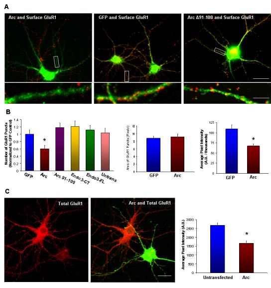
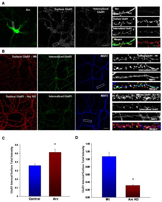
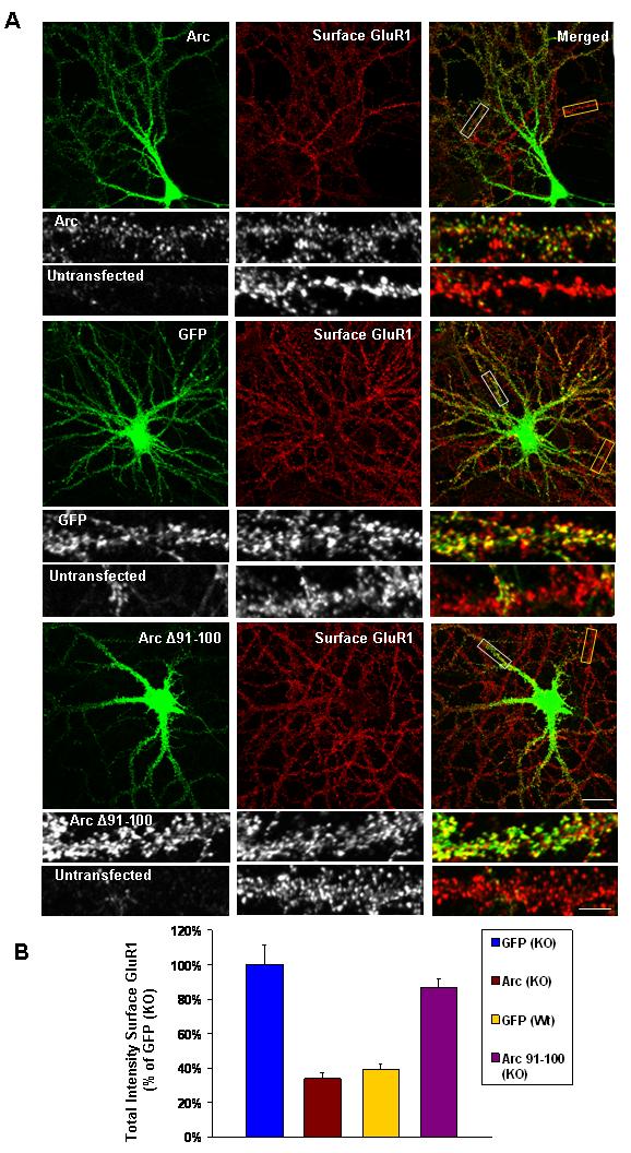
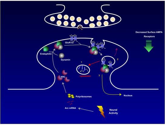
Arc/Arg3.1 is an immediate-early gene whose mRNA is rapidly transcribed and targeted to dendrites of neurons as they engage in information processing and storage. Moreover, Arc/Arg3.1 is known to be required for durable forms of synaptic plasticity and learning. Despite these intriguing links to plasticity, Arc/Arg3.1's molecular function remains enigmatic. Here, we demonstrate that Arc/Arg3.1 protein interacts with dynamin and specific isoforms of endophilin to enhance receptor endocytosis. Arc/Arg3.1 selectively modulates trafficking of AMPA-type glutamate receptors (AMPARs) in neurons by accelerating endocytosis and reducing surface expression. The Arc/Arg3.1-endocytosis pathway appears to regulate basal AMPAR levels since Arc/Arg3.1 KO neurons exhibit markedly reduced endocytosis and increased steady-state surface levels. These findings reveal a novel molecular pathway that is regulated by Arc/Arg3.1 and likely contributes to late-phase synaptic plasticity and memory consolidation.


Figures

Figure 1
Figure 1
Arc Interacts with Endophilin and Dynamin(A) Schematic diagram showing the domain structure of Arc, endophilin 3, dynamin 2, and the yeast two hybrid fragments of each. (B) When co-expressed in HEK293 cells Arc co-IPs dynamin 2(503-871) but not dynamin 2(612-871) (lacks the PH domain). Control blots show expressed dynamin (dynamin Off) and immunoprecipitated Arc (Arc IP). (C) Dynamin co-IPs with Arc from a rat forebrain synaptosomal fraction (P2). 10% of lysate used for IP was routinely loaded in offered lane. (D) Arc co-IPs endophilin 2(172-368) and endophilin 3(172-347), but not endophilin 1(172-352) from HEK293 cells. (E) Pan endophilin Ab detects co-IP with Arc from rat forebrain P2 fraction. Samples were reblotted with Abs selective for individual endophilin A proteins. Arc IP fractions show enrichment for endophilin 2 and 3, but not endophilin 1.
Figure 2
Figure 2
Endophilin 2, 3 and Arc Localize to Post-synaptic Sites with GluR1 in Hippocampal Neurons (A)Immunogold localization of endophilin 1 (a-d), 2 (e-h) and 3 (i-m) in the hippocampus CA1 stratum radiatum (a,b, e,f, i,j,l) and molecular layer of the dentate gyrus (c,d,g,h,k,m). Arrows indicate clusters of gold in the postsynaptic spine (f-k) or dendrite shaft (g-right, l,m). This labeling often is associated with vesicular or tubulovesicular structures (arrows in f-h,j,l,m). Labeling for endophilin 1 is common mainly in presynaptic terminals (pre). Endophilin 2 labeling also is common in presynaptic terminals as well as in postsynaptic spines. Endophilin 3 labeling is seen less commonly in presynaptic terminals (compared to endophilin 1 and 2) but is prominent in postsynaptic spines (similar to endophilin 2). Scale bar is 100 nm. Results from the counting of 180 synapses (from random sections) of the hippocampus CA1 stratum radiatum are as follows. For endophilin 1, there were 0.483 gold/postsynaptic spine and 1.933 gold/presynaptic terminal, so that there were 4x as many gold particles in the pre as in the post-synapse. The difference is highly significant (p≪0.001). For endophilin 2, there were 1.283 gold/postsynaptic spine and 2.056 gold/presynaptic terminal, so that there were 1.6x as many gold particles in the pre as in the post-synapse. The difference is highly significant (p<0.001). For endophilin 3, there were 0.367 gold/postsynaptic spine and 0.422 gold/presynaptic terminal, so that there were 1.2x (essentially 1x) as many gold particles in the pre as in the post-synapse. (B) Immuno EM images of CA1 region of adult rat brain showing Arc localization in the postsynaptic density (PSD). Bottom panel shows Arc immunoreactivity at the lateral margin of the PSD. (C) Immunostaining of 4 week old hippocampal cultured neurons shows endogenous Arc and endophilin 3 localized at dendrites, and in synapses marked by GluR1 (yellow arrows). Some Arc/endophilin 3/GluR1 puncta colocalize together at non-synaptic sites (white arrows) in the dendritic shaft. (Scale dendrite)
Figure 3
Figure 3
Arc is Recruited with Endophilin and Dynamin to Vesicles in HeLa Cells(A) Arc expressed alone has a cytoplasmic and nuclear distribution in HeLa cells, with some enrichment at the plasma membrane. En3-CT [Endophilin 3(172-347)] alone has a cytoplasmic distribution with enrichment at the plasma membrane. Dyn2-CT [Dynamin 2(503-871)] alone localizes to vesicular structures near the plasma membrane. When Arc and En3-CT are expressed together, vesicular structures form that contain both proteins. Arc and Dyn2-CT co-expression results in a redistribution of Arc to dynamin 2 containing vesicular structures. When Arc, Dyn2-CT, and En3-FL (endophilin 3 full length) are co-expressed, all three proteins colocalize in vesicular structures. (B) Arc and full-length endophilin 3 have no effect on Alexa-555 conjugated transferrin uptake when expressed alone (a and c). Arc and myc antibodies were used to stain Arc and endophilins respectively. (b) En3-CT inhibits transferrin uptake. (d) Arc and En3-CT co-expression results in endosomes that internalize transferrin. (Scale bars represent 30 μm)
Figure 4
Figure 4
Distinct Regions of Arc Bind to Endophilin and Dynamin(A) Arc or Arc (1-154) co-IP endophilin 3(172-347), while Arc (155-396) does not. (B) The region of Arc that binds endophilin maps to aa 81-130. Internal deletion of 50-80 does not reduce binding to endophilin 3(172-347), while deletions in the region 81-130 do. Non-specific bands are indicated with *. (C) GST-Endophilin 3(172-347) binds Arc expressed in HEK293 cells, and the interaction is blocked by peptide Arc 89-100, but not by Arc 76-88 or an unrelated peptide (Homer ligand). Final concentration of peptides is 100μM. (D)Arc (155-396) co-IPs with dynamin 2(503-871), while Arc (1-154) does not. (E) Internal deletions of 20 aa that span the region Arc (195-214) disrupt co-IP with Dynamin 2(503-871) co-expressed in HEK293 cells. No change in binding with the deletion of region Arc(235-254). (F) Arc co-IPs endophilin 3(172-347) more effectively than endophilin 3 (En3-FL) and is not altered by SH3 mutation [W322A]. (G)Deletions of BAR domain modify co-IP with Arc. (H)GST-Endophilin 2 (218-254) binds Arc.
Figure 5
Figure 5
Arc and Endophilin Associate with Endosomes in Neurons(A)Arc and endophilin 3 transgenes co-localize in large puncta in the dendritic shaft and in spines. (B) Neurons were incubated with a GluR1 N-terminal antibody at 10°C, and were subsequently allowed to undergo basal endocytosis for 30 min at 37°C. Any remaining surface GluR1 were stripped with an acid wash. Internalized GluR1 puncta co-localize with many of the endophilin-Arc transgene vesicular puncta. (C) Neurons were incubated with Alexa555-transferrin for one hour at 37°C, labeling both early and recycling endosomal pools. A number of Arc-endophilin transgene puncta co-localized with transferrin in dendrites (yellow arrows).
Figure 6
Figure 6
Arc Expression Decreases Surface and Total AMPARs(A) Representative images of Arc transgene expression (green), showing a loss of surface GluR1 puncta (assessed using an N-terminal antibody, (red) as compared to neighboring untransfected cells, 16 hours post-transfection. GFP transfected neurons had normal numbers of-100 expression GluR1 had no puncta. effect on the number of GluR1 puncta. (B) Arc expression causes a significant decrease in the number of surface GluR1 puncta (n = 48 dendrites from16 cells, *p < 0.01) compared with neighboring untransfected cells. GFP transfection had no effect. In contrast,-100, endophilin Arc 3(172-347) or endophilin 3 had no significant effect on GluR1 puncta. Arc expression did not change GluR1 puncta size or average pixel intensity, but total intensity was significantly decreased *p < 0.001). (C) Arc transgene reduced total GluR1 (assessed using a C-terminal antibody), as compared to untransfected neurons 16 hours post-transfection (n = 60 regions from 20 cells, *p < 0.001).
Figure 7
Figure 7
The Arc-Dynamin Interaction is Required for the Arc-dependent Decrease in Surface AMPARs. (A) Arc and dynamin2-CT (aa503-871) transgenes co-localize in large puncta in the dendritic shaft and in spines. (B) Representative images of Arc transgene expression, showing a loss of surface GluR1 puncta/intensity, whereas Arc-214 Δ195 expression had no effect on the number of GluR1 puncta/intensity. Quantition shows that Arc expression causes a significant decrease in the number and total intensity of surface GluR1 puncta (n = 63/21). In contrast,-214, Arc had no significant Δ195effect on GluR1 puncta or total intensity (n = 57/10). (*p < 0.001).
Figure 8
Figure 8
Arc Increases AMPAR Endocytosis while Arc KO Neurons Exhibit Increased Surface AMPARs and a Deficit in Endocytosis (A)Arc transfected neuron with both surface and internalized GluR1 images using different secondary antibodies after 30 min basal endocytosis. Magnified dendrites show an Arc transfected dendrite and an untransfected dendrite. (B) Representative images of surface and internalized GluR1 after 30 min of basal endocytosis in Wt and KO hippocampal neurons. MAP2 staining is shown to highlight individual dendrites (Scale bar, 30μm) (C) Quantification of surface and internalized GluR1. Arc expression results in a decrease in the surface to internalized GluR1 ratio (n = 30/10, p < 0.02), indicating that more endocytosis of receptors occurred in Arc transfected neurons. Similar results are reported in Figure 3 of Shepherd et al (this issue), although the results reported reflect two different experiments. (*p < 0.05). (D)Total intensity of surface and internalized GluR1 puncta was quantified to assess the amount of endocytosis during 30 min. Arc KO neurons (n = 48/16) have a significantly higher surface to internalized ratio than Wt neurons (n = 51/17). (*p ≪ 0.001).
Figure 9
Figure 9
Arc Expression in Arc KO Neurons Rescues Surface GluR1 Levels (A)Representative images of Arc Sindbis virus expression in Arc KO low-density hippocampal neurons, showing a reduction of surface GluR1 as compared to neighboring untransfected cells. GFP Sindbis virus expression has no affect on surface GluR1. In contrast to-100 Arc Sindbis virus expression does not affect surface GluR1 levels. (White boxes show magnified transfected dendrites and yellow boxes highlight untransfected dendrites. Scale bars, 30μm and 8 μm in magnified dendrites) (B)Quantification of surface GluR1 experiments. Arc expression in Arc KO neurons (n = 30/10) significantly reduces total intensity of surface GluR1 (34 ± 3% of GFP transfected KO neurons, n = 30/10, p < 0.001) and is comparable to GluR1 levels in-100 expression in Arc KO neurons does not rescue GluR1 levels (87 ± 5%, n = 30/10, p = 0.3)
Figure 10
Figure 10
Proposed Model for Arc Modulation of AMPAR TraffickingArc mRNA is induced after neuronal activity and is transported to dendrites. Upregulation of Arc protein occurs in a synapse-specific manner via local translation. Arc protein recruits endophilin and dynamin, which is anchored to the PSD via its interaction with VSCC (Chen et al., 2003) and Shank (Okamoto et al., 2001), respectively. The complex of Arc, endophilin and dynamin modulates endosome formation, and selectively recruits AMPA receptors. These endosomes may then 1. Recycle back to the membrane. 2. Traffic to the lysosomal compartment where protein is degraded or sent to the proteosome 3. Traffic to the nucleus as a signaling endosome carrying other cargo. It is interesting to note that Arc protein is present in the nucleus of select populations of brain neurons where it is coincidently induced with Arc protein in dendrites. Changes in the kinetics of these vesicular pathways are anticipated to underlie changes in steady state levels of AMPAR at synapses.

Similar articles

Cited by

References

    1. Banker GA, Cowan WM. Rat hippocampal neurons in dispersed cell culture. Brain Res. 1977;126:397–342. - PubMed
    1. Barik S. Megaprimer PCR. Methods Mol Biol. 2002;192:189–196. - PubMed
    1. Blanpied TA, Scott DB, Ehlers MD. Dynamics and regulation of clathrin coats at specialized endocytic zones of dendrites and spines. Neuron. 2002;36:435–449. - PubMed
    1. Bock J, Thode C, Hannemann O, Braun K, Darlison MG. Early socio-emotional experience induces expression of the immediate-early gene Arc/arg3.1 (activity-regulated cytoskeleton-associated protein/activity-regulated gene) in learning-relevant brain regions of the newborn chick. Neuroscience. 2005;133:625–633. - PubMed
    1. Boehm J, Malinow R. AMPA receptor phosphorylation during synaptic plasticity. Biochem Soc Trans. 2005;33:1354–1356. - PubMed

Publication types

MeSH terms