Comparative Study
. 2006 Dec;15(12):2697-707.
doi: 10.1110/ps.062398606. Epub 2006 Nov 6.

An inserted Gly residue fine tunes dynamics between mesophilic and thermophilic ribonucleases H

Affiliations
Comparative Study

An inserted Gly residue fine tunes dynamics between mesophilic and thermophilic ribonucleases H

Joel A Butterwick et al. Protein Sci. 2006 Dec.

Abstract

Dynamic processes are inherent properties of proteins and are crucial for a wide range of biological functions. To address how changes in protein sequence and structure affect dynamic processes, a quantitative comparison of microsecond-to-microsecond time scale conformational changes, measured by solution NMR spectroscopy, within homologous mesophilic and thermophilic ribonuclease H (RNase H) enzymes is presented. Kinetic transitions between the observed major state (high population) and alternate (low population) conformational state(s) of the substrate-binding handle region in RNase H from the mesophile Escherichia coli (ecRNH) and thermophile Thermus thermophilus (ttRNH) occur with similar kinetic exchange rate constants, but the difference in stability between exchanging conformers is smaller in ttRNH compared to ecRNH. The altered thermodynamic equilibrium between kinetically exchanging conformers in the thermophile is recapitulated in ecRNH by the insertion of a Gly residue within a putative hinge between alpha-helices B and C. This Gly insertion is conserved among thermophilic RNases H, and allows the formation of additional intrahelical hydrogen bonds. A Gly residue inserted between alpha-helices B and C appears to relieve unfavorable interactions in the transition state and alternate conformer(s) and represents an important adaptation to adjust conformational changes within RNase H for activity at high temperatures.


Figures

Figure 1.
Figure 1.
Sequence and structural alignments of RNases H. (A) ClustalW (Thompson et al. 1994) multiple sequence alignment of RNase H from the mesophilic bacterium E. coli (37°C) and several thermophilic bacteria: Chlorobium tepidum (47°C), Moorella thermoacetica (58°C), T. thermophilus (70°C), and Thermoanaerobacter tengcongensis (75°C). The optimal growth temperature for each organism is indicated in parentheses. Conserved active site and substrate-binding residues are highlighted in blue; the inserted Gly is shown in red. α-Helical (αA–αE) and β-strand (β1–β5) secondary structural elements and the handle in WT ecRNH are indicated above the alignment. (B) Structural superposition of WT ecRNH (blue, PDB 2RN2), WT ttRNH (orange, PDB 1RIL), and iG80b ecRNH (gray, PDB 1GOA) using backbone atoms along core helices αA and αD. Structural elements from (A) and the N and C termini are indicated. Ribbon diagrams for this and subsequent figures were drawn using MOLMOL (Koradi et al. 1996).
Figure 2.
Figure 2.
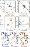
Structural consequences of the Gly insertion. (A) Chemical shift changes between WT and iG80b ecRNH (Δδec) and WT and dG85 ttRNH (Δδtt) are compared as Δδ = δmutant−δWT for backbone amide (left) 1H and (right) 15N nuclei. Residues in αB, αC and the handle (70–100 in ecRNH) are indicated by open circles; the correlation coefficient for these residues are −0.63 for 1H and −0.84 for 15N. (B) Overlay of 1H–15N correlation spectra for iG80b ecRNH and dG85 ttRNH with (left) WT ttRNH and (right) WT ecRNH highlighting the positions of Thr79 (ecRNH) and Thr83 (ttRNH). (C) Main-chain hydrogen bonds in the αBC hinge and handle are shown in green for WT ecRNH (blue, PDB 2RN2), iG80b ecRNH (gray, PDB 1GOA), and WT ttRNH (orange, PDB 1RIL). Additional hydrogen bonds seen in the crystal structures of iG80b ecRNH and WT ttRNH are shown in magenta.
Figure 3.
Figure 3.
Chemical exchange line broadening in WT and mutant RNases H. Backbone amide 15N R ex at 300 K, 14.1 T for WT ecRNH, WT ttRNH, iG80b ecRNH, and dG85 ttRNH are shown. Residues with significant chemical exchange line broadening (R ex ≥ 2.5 sec−1) are indicated by open circles. Vertical dashed lines indicate the sites of mutation. Secondary structural elements are diagrammed at the top of the figure. Data for WT ttRNH are from Butterwick et al. (2004).
Figure 4.
Figure 4.
Temperature dependence of chemical exchange line broadening. (A) R ex measured at multiple temperatures and 14.1 T and (B) Arrhenius plots are shown for (left) Trp90 in WT and iG80b ecRNH and (right) Trp95 in WT and dG85 ttRNH. Open and closed symbols represent data for WT and mutant RNases H, respectively. Apparent activation energies, Ê a, derived from the slope of the lines in B are listed in Table 1. Square symbols represent data that deviate from a linear Arrhenius relationship, and are not included for determining Ê a. Data for WT ttRNH are from Butterwick et al. (2004).
Figure 5.
Figure 5.
R relaxation dispersion for backbone amide 15N nuclei. R 2(ω e) dispersion results are shown for residues Trp90 and Lys60 in WT and iG80b ecRNH, and Trp95 and Val126 in WT ttRNH at 300 K. Closed and open symbols represent data collected at 11.7 and 18.8 T, respectively.
Figure 6.
Figure 6.
Summary of conformational changes in RNases H. Energy diagram comparing the relative stabilities and activation energies for exchanging conformers in WT ecRNH (blue), WT ttRNH (orange), and iG80b ecRNH (gray) at 300 K. Diagram is drawn to scale assuming p B = 1% for WT ecRNH and k o = k B T/h, where k B and h are Boltzmann's and Planck's constants, respectively.

Similar articles

Cited by

References

    1. Akke, M. 2002. NMR methods for characterizing microsecond to millisecond dynamics in recognition and catalysis. Curr. Opin. Struct. Biol. 12: 642–647. - PubMed
    1. Akke, M. and Palmer III, A.G. 1996. Monitoring macromolecular motions on microsecond to millisecond time scales by R 1ρ − R 1 constant relaxation time NMR spectroscopy. J. Am. Chem. Soc. 118: 911–912.
    1. Benkovic, S.J. and Hammes-Schiffer, S. 2003. A perspective on enzyme catalysis. Science 301: 1196–1202. - PubMed
    1. Butterwick, J.A., Loria, J.P., Astrof, N.S., Kroenke, C.D., Cole, R., Rance, M., and Palmer III, A.G. 2004. Multiple time scale backbone dynamics of homologous thermophilic and mesophilic ribonuclease HI enzymes. J. Mol. Biol. 339: 855–871. - PubMed
    1. Cavanagh, J., Fairbrother, W.J., Palmer III, A.G., and Skelton, N.J. 1996. Protein NMR spectroscopy: Principles and practice. Academic Press, San Diego, CA.

Publication types

LinkOut - more resources