. 2006 Nov 6;175(3):369-75.
doi: 10.1083/jcb.200511112.

A new method reveals microtubule minus ends throughout the meiotic spindle

Affiliations

A new method reveals microtubule minus ends throughout the meiotic spindle

Kendra S Burbank et al. J Cell Biol. .

Abstract

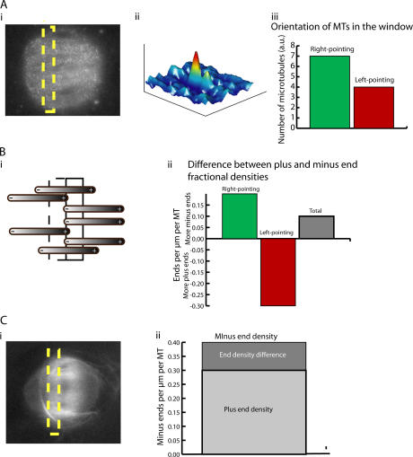
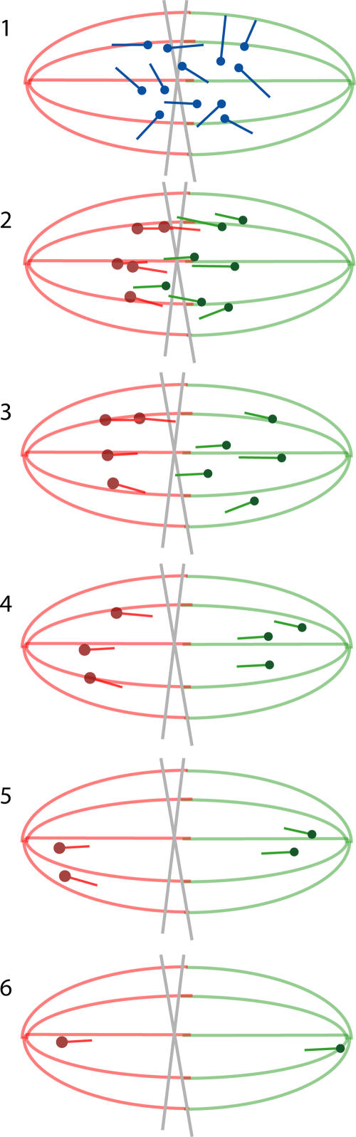
Anastral meiotic spindles are thought to be organized differently from astral mitotic spindles, but the field lacks the basic structural information required to describe and model them, including the location of microtubule-nucleating sites and minus ends. We measured the distributions of oriented microtubules in metaphase anastral spindles in Xenopus laevis extracts by fluorescence speckle microscopy and cross-correlation analysis. We localized plus ends by tubulin incorporation and combined this with the orientation data to infer the localization of minus ends. We found that minus ends are localized throughout the spindle, sparsely at the equator and at higher concentrations near the poles. Based on these data, we propose a model for maintenance of the metaphase steady-state that depends on continuous nucleation of microtubules near chromatin, followed by sorting and outward transport of stabilized minus ends, and, eventually, their loss near poles.


Figures

Figure 1.
Figure 1.
Schematic of method for determining the fractional densities of plus and minus ends in a portion of the spindle. (A) Measurement of the density of left- and right-pointing MTs. (i) Cross-correlation measures flow of speckles within the window (dashed box). The two peaks represent the flow toward each amount of the two poles. (ii) The volume of each peak gives the number of microtubules in the window fluxing in each direction. MTs moving left are assumed to have plus ends pointing right. (iii) Orientation of MTs in the window. (B) Calculation of the difference between fractional densities of plus and minus ends. (i) The number of right-pointing MTs is compared with the number in the adjacent window to the right. The change measures the difference in the number of plus versus minus ends in the window. This is because MTs with no ends in the two windows extend through both. MTs with plus ends extend only through the left window, while those with minus ends only through the right window. The difference between fractional densities of minus versus plus ends for left-pointing MTs is calculated in a similar manner, by comparing the number of left-pointing MTs with the number in the window to the left. The total difference is the sum of the left- and right-pointing differences. (ii) Difference between plus and minus end fractional densities. (C) Measurement of the density plus ends and calculation of the minus end fractional density. New tubulin incorporates into growing plus ends. The total number of plus ends in the window is found by measuring how quickly fluorescence intensity increases immediately after the addition of labeled tubulin. The fractional density of plus ends is added to the difference between plus and minus end fractional densities. This gives the fractional density of minus ends in the window. (ii) Fractional density of minus ends in the window.
Figure 2.
Figure 2.
Oriented MT number distributions. Plots for 14 spindles showing oriented MT number distribution, in arbitrary units, versus position along the spindle-pole axis. Distributions for right-pointing MTs are given in solid lines, and distributions for left-pointing MTs are given in dotted lines. Spindle-pole positions, which were manually selected by the edge of visible florescence in spindle images, are marked with vertical dashed lines. The distance between tick marks on the x axis is 10 μm.
Figure 3.
Figure 3.
Plus and minus end density distributions. (A) A gallery of minus- (solid) and plus-end (dotted) fractional density distributions versus position along spindle-pole axis. The distance between tick marks on the x axis is 10 μm. Tick marks on the y axis represent 0.05 ends/μm, and dashed lines are at 0 ends/μm. (B) Plots of minus (solid) and plus end (dotted) density distributions, for the same spindles shown in A. X ticks represent 10 μm, and the y axes are in arbitrary units with dashed lines at 0.
Figure 4.
Figure 4.
Model of steady-state spindle formed by chromosomal nucleation and stochastic MT loss. A steady-state spindle might be maintained through chromosomal nucleation, poleward sliding of MTs caused by the flux mechanism, and MT slowdown upon approach to the poles. (1) MTs are nucleated in a region around the chromosomes. (2) They initially point in random directions, but are sorted by motors to be parallel with the dominant MT orientation at the point of nucleation. (3–6) The MTs are moved poleward by a flux mechanism. Throughout the process, MTs and their minus ends disappear stochastically. As MTs disappear, new ones are continuously nucleated near the center.

Similar articles

Cited by

References

    1. Brinkley, B.R. 1985. Microtubule organizing centers. Annu. Rev. Cell Biol. 1:145–172. - PubMed
    1. Carazo-Salas, R.E., and E. Karsenti. 2003. Long-range communication between chromatin and microtubules in Xenopus egg extracts. Curr. Biol. 13:1728–1733. - PubMed
    1. Carazo-Salas, R.E., G. Guarguaglini, O.J. Gruss, A. Segref, E. Karsenti, and I.W. Mattaj. 1999. Generation of GTP-bound Ran by RCC1 is required for chromatin-induced mitotic spindle formation. Nature. 400:178–181. - PubMed
    1. Carazo-Salas, R.E., O.J. Gruss, I.W. Mattaj, and E. Karsenti. 2001. Ran-GTP coordinates regulation of microtubule nucleation and dynamics during mitotic-spindle assembly. Nat. Cell Biol. 3:228–234. - PubMed
    1. Desai, A., A. Murray, T.J. Mitchison, and C.E. Walczak. 1999. The use of Xenopus egg extracts to study mitotic spindle assembly and function in vitro. Methods Cell Biol. 61:385–412. - PubMed

Publication types