. 2006 Nov 6;175(3):477-90.
doi: 10.1083/jcb.200602085.

Reassembly of contractile actin cortex in cell blebs

Affiliations

Reassembly of contractile actin cortex in cell blebs

Guillaume T Charras et al. J Cell Biol. .

Abstract

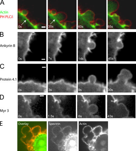
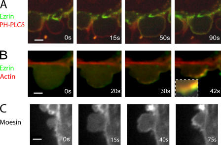
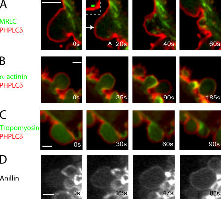
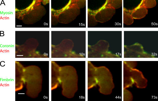
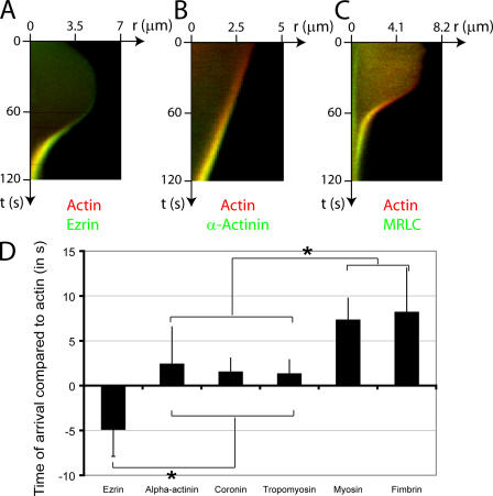
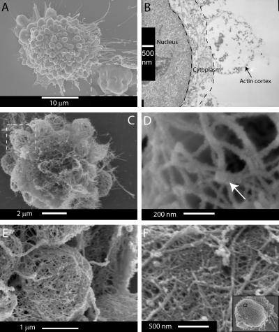
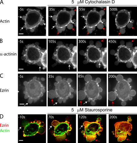
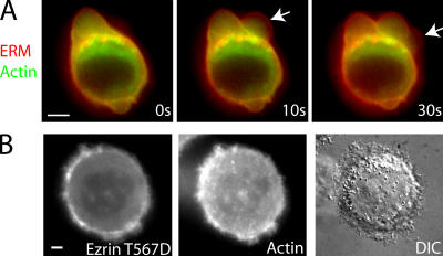
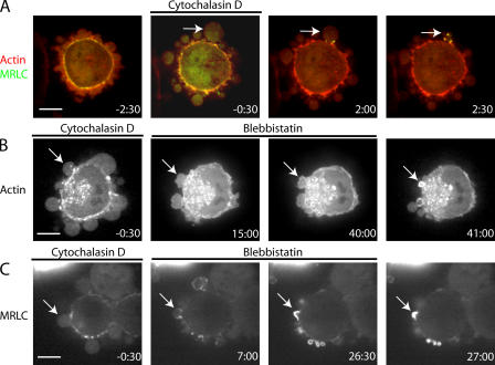
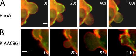
Contractile actin cortex is involved in cell morphogenesis, movement, and cytokinesis, but its organization and assembly are poorly understood. During blebbing, the membrane detaches from the cortex and inflates. As expansion ceases, contractile cortex re-assembles under the membrane and drives bleb retraction. This cycle enabled us to measure the temporal sequence of protein recruitment to the membrane during cortex reassembly and to explore dependency relationships. Expanding blebs were devoid of actin, but proteins of the erythrocytic submembranous cytoskeleton were present. When expansion ceased, ezrin was recruited to the membrane first, followed by actin, actin-bundling proteins, and, finally, contractile proteins. Complete assembly of the contractile cortex, which was organized into a cagelike mesh of filaments, took approximately 30 s. Cytochalasin D blocked recruitment of actin and alpha-actinin, but had no effect on membrane association of ankyrin B and ezrin. Ezrin played no role in actin nucleation, but was essential for tethering the membrane to the cortex. The Rho pathway was important for cortex assembly in blebs.


Figures

Figure 1.
Figure 1.
Localization of proteins during bleb expansion and retraction. All images were acquired using confocal microscopy. Timing relative to the first image is indicated in white text. (A) Localization of the membrane marker (the PH domain of PLCδ, red) and actin (green) during bleb expansion and retraction. The actin cortex is intact during bleb expansion (arrows) and is disassembled at later time points. (B–D) Ankyrin B (B), protein 4.1 (C), and myosin I (myr3, D) localize to the cell membrane throughout the life of a bleb. (E) Spectrin (green) colocalizes with actin (red) in retracting blebs. Bars, 1 μm.
Figure 2.
Figure 2.
Localization of ERM proteins during bleb expansion and retraction. All images were acquired using confocal microscopy. Timing relative to the first image is indicated in white letters. (A) Ezrin (green) is initially absent from the membrane of expanding blebs. Once expansion stops, ezrin is recruited to the bleb and forms a continuous rim colocalized with the membrane. The bleb membrane (red) is visualized using the PH domain of PLCδ. (B) Ezrin (green) appears before actin (at 20 vs. 30 s) and forms a continuous rim external to the actin shell (red, inset). (C) Moesin is absent from the membrane of expanding blebs, but later forms a continuous rim. Bars, 2 μm.
Figure 3.
Figure 3.
Localization of proteins relative to the membrane during bleb retraction. All images were acquired using confocal microscopy. The PH domain of PLCδ, a membrane marker, is visualized in red and the protein of interest in green. Timing relative to the first image is indicated in white text. MRLC (A) localizes to distinct foci (arrows and inset) under the bleb membrane in retracting blebs. α-Actinin (B), tropomyosin (C), and anillin (D) all form a continuous rim under the bleb membrane once expansion has stopped. Bars, 2 μm.
Figure 4.
Figure 4.
Localization of proteins relative to actin during bleb retraction. All images were acquired using confocal microscopy. In all images, actin is in red and the protein of interest in green. Timing relative to the first image is indicated in white text. (A) MRLC is originally not present (0–15 s) in the bleb, but assembles in distinct foci underneath the actin shell. (B) Coronin appears shortly after actin and forms a continuous rim colocalized with the actin shell. (C) Fimbrin appears after actin (44 vs. 18 s) and forms a continuous rim colocalized with the actin shell. Bars, 2 μm.
Figure 5.
Figure 5.
Timing of arrival of actin binding proteins in the bleb relative to actin. (A–C) Kymographs showing the localization of actin (red) compared with actin-binding proteins (green) during bleb retraction. Bleb extension is shown on the horizontal axis and time is shown on the vertical axis. Ezrin (A, green) arrives in the bleb significantly before actin and, consequently, a green trace can be seen at the bleb rim. As actin gets recruited to the rim, the trace turns yellow because of colocalization with ezrin. α-Actinin (B, green) and MRLC (C, green) are recruited after actin (red) and, consequently, a red trace can be seen first at the bleb rim. This trace turns yellow as these proteins are recruited to the rim. (D) Timing of arrival of the different actin-binding proteins in relationship to actin (t = 0 s). The error bars represent SD. Asterisks denote significant differences in timing. Ezrin arrives at the bleb rim significantly before actin. Coronin, α-actinin, and tropomyosin arrive significantly later than actin. Finally, fimbrin and myosin II appear last.
Figure 6.
Figure 6.
Actin ultrastructure in retracting blebs. (A) SEM of a blebbing cell with an intact cell membrane. (inset) As the retraction ends, the bleb membrane is crumpled. (B) TEM of the actin cortex of a retracting bleb. The bleb interior is devoid of cytoskeletal structures. Actin is concentrated at the bleb rim and forms a thin shell that is 10–20 nm thick. (C) Actin cytoskeleton of a blebbing cell. (D) Enlargement of boxed area in C. The cortex is formed by an entangled meshwork of actin with a few protuberant knots (arrow). The mesh size is ∼200 nm. Myosin S1 head decoration shows no specific orientation of the actin around the knots. (E) Actin cytoskeleton of a bleb in a dividing HeLa cell. The morphology of blebs is similar to that of filamin-deficient cells, but the mesh is finer. (F) Actin cortex of a rounded HeLa cell. The morphology of the cortex is similar to that of blebs. The entire cell is shown in the inset.
Figure 7.
Figure 7.
Localization of actin and actin-binding proteins during bleb expansion and retraction in the presence of drugs. Cytochalasin D, an actin depolymerizer, was added at time t = 0 s. All images were acquired using confocal microscopy. The timing of each image is indicated in white text. In all images, white arrows indicate blebs that have formed before the addition of cytochalasin D and red arrows indicate blebs that emerge after cytochalasin D treatment. (A and B) Actin (A) and α-actinin (B) localize to the rim of blebs that have formed before treatment and which continue to retract. Blebs that form after treatment do not retract and the proteins stay cytoplasmic. (C) Ezrin localizes to the rim of blebs that have formed before treatment, as well as to the rim of those formed after treatment. (D) In the absence of staurosporine, ezrin localizes to the bleb rim (white arrow, t = −10 s). Blebs that form after the addition of staurosporine (t = 0 s), do not recruit ezrin to their rim, but do still form an actin rim (red arrows). Treatment with staurosporine causes the relocalization of extant ezrin from the bleb rim to the cytoplasm (white arrows). Bars, 5 μm.
Figure 8.
Figure 8.
Mutant forms of ezrin modulate the attachment of the cell membrane to the actin cytoskeleton. All images were acquired using wide-field microscopy. Timing relative to the first image is indicated in white letters. (A) Microinjection of the FERM domain of ezrin tagged with mRFP into cells expressing actin-GFP fragilizes the attachment of the membrane to the actin cortex. In retracting blebs, the actin rim tended to tear away from the membrane (arrows at times t = 10 s and t = 30 s). This resulted in inwards flow of the actin rim, but the membrane failed to retract. (B) Microinjection of constitutively active ezrin (ezrin T567D) tagged with GFP into actin-mRFP–expressing cells causes the cessation of blebbing. Ezrin T567D colocalizes with actin at the cell periphery. Bars, 5 μm.
Figure 9.
Figure 9.
Myosin II powers bleb retraction cytochalasin D, an actin depolymerizer, was added at time t = −2 min and treatment was pursued until t = 0 min. Cytochalasin was washed out and, in some cases, blebbistatin was included in the washout medium. This second treatment was pursued for 15–50 min, and then a final washout or inactivation was effected. All images were acquired using confocal microscopy. The timing of each image in minutes is indicated in white text. (A) Blebs that emerged after the onset of treatment formed actin puncta (red), recruited MRLC (green) to puncta, and retracted upon washout (white arrow). (B) Inclusion of the myosin ATPase inhibitor blebbistatin in the washout medium did not prevent the formation of an actin rim but stopped retraction (white arrow). Upon blebbistatin inactivation (t = 50 min), retraction proceeded normally. (C) Inclusion of blebbistatin in the washout medium did not inhibit myosin recruitment to the bleb rim (white arrow). Upon blebbistatin inactivation (t = 27 min), retraction proceeded normally. Bars, 5 μm.
Figure 10.
Figure 10.
Localization of RhoGTPase RhoA and the RhoGEF KIAA0861 during blebbing. Timing relative to the first image is indicated in white text. All images were acquired using confocal microscopy. In all images, actin is shown in green and the protein of interest in red. Both RhoA (A) and the RhoGEF KIAA0861 (B) localized to the bleb membrane during all phases of blebbing. In both cases, the proteins were displaced toward the cell exterior when compared with actin. Bars, 2 μm.

References

    1. Alberts, B., D. Bray, J. Lewis, M. Raff, K. Roberts, and J. Watson. 2004. Molecular Biology of the Cell. 4th edition. Taylor and Francis, New York. 1463 pp.
    1. Barylko, B., D.D. Binns, and J.P. Albanesi. 2000. Regulation of the enzymatic and motor activities of myosin I. Biochim. Biophys. Acta. 1496:23–35. - PubMed
    1. Bement, W.M., H.A. Benink, and G. von Dassow. 2005. A microtubule-dependent zone of active RhoA during cleavage plane specification. J. Cell Biol. 170:91–101. - PMC - PubMed
    1. Bennett, V., and A.J. Baines. 2001. Spectrin and ankyrin-based pathways: metazoan inventions for integrating cells into tissues. Physiol. Rev. 81:1353–1392. - PubMed
    1. Bray, D., and J.G. White. 1988. Cortical flow in animal cells. Science. 239:883–888. - PubMed

Publication types

MeSH terms