. 2006 Nov 6;175(3):491-503.
doi: 10.1083/jcb.200607036.

A postsynaptic spectrin scaffold defines active zone size, spacing, and efficacy at the Drosophila neuromuscular junction

Affiliations

A postsynaptic spectrin scaffold defines active zone size, spacing, and efficacy at the Drosophila neuromuscular junction

Jan Pielage et al. J Cell Biol. .

Abstract

Synaptic connections are established with characteristic, cell type-specific size and spacing. In this study, we document a role for the postsynaptic Spectrin skeleton in this process. We use transgenic double-stranded RNA to selectively eliminate alpha-Spectrin, beta-Spectrin, or Ankyrin. In the absence of postsynaptic alpha- or beta-Spectrin, active zone size is increased and spacing is perturbed. In addition, subsynaptic muscle membranes are significantly altered. However, despite these changes, the subdivision of the synapse into active zone and periactive zone domains remains intact, both pre- and postsynaptically. Functionally, altered active zone dimensions correlate with an increase in quantal size without a change in presynaptic vesicle size. Mechanistically, beta-Spectrin is required for the localization of alpha-Spectrin and Ankyrin to the postsynaptic membrane. Although Ankyrin is not required for the localization of the Spectrin skeleton to the neuromuscular junction, it contributes to Spectrin-mediated synapse development. We propose a model in which a postsynaptic Spectrin-actin lattice acts as an organizing scaffold upon which pre- and postsynaptic development are arranged.


Figures

Figure 1.
Figure 1.
Postsynaptic elimination of α- and β-Spectrin at the NMJ. (A–D) A third instar wild-type synapse at muscle 4 is stained for α-Spectrin (A), β-Spectrin (B), and the presynaptic membrane marker HRP (D). α- and β-Spectrin colocalize and are highly enriched in the postsynaptic SSR (C). (E–H) Muscle-specific expression of α-spectrin dsRNA leads to the elimination of α-Spectrin in the muscle, but not in the motoneuron axon (E). β-Spectrin protein levels in the muscle are unchanged, but the distribution of β-Spectrin within the SSR is severely affected (F and G). (I–L) Muscle-specific expression of β-spectrin dsRNA results in the elimination of β- and α-Spectrin from the postsynaptic muscle (I–K). The presynaptic nerve terminal can be identified by HRP (L). A second instar wild-type synapse is shown stained for β-Spectrin (M and N) and HRP (N). (O and P) Expression of β-spectrin dsRNA results in the elimination of β-Spectrin from the muscle in second instar larvae. (Q) Western blot analysis of third instar larvae. The ubiquitous expression of α-spectrin dsRNA results in the elimination of α-Spectrin, but not β-Spectrin protein levels. The ubiquitous expression of β-spectrin dsRNA greatly reduces β-Spectrin protein, with only minor effects on α-Spectrin protein levels. Bars, (A–P) 10 μm.
Figure 2.
Figure 2.
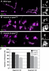
Loss of postsynaptic α- or β-Spectrin results in severe morphological defects in NMJ formation. (A–C) NMJs from muscle 6 and 7 at segment A3 are shown stained for the presynaptic vesicle marker synaptotagmin (Syt; green) and for the postsynaptic SSR marker Discs-large (Dlg; magenta). (right) Higher resolution images of areas indicated by asterisks. (A) In wild type, the postsynaptic marker Dlg tightly surrounds the presynaptic nerve terminal marked by Syt. (B) A NMJ lacking postsynaptic α-Spectrin. NMJ size is reduced and the number of synaptic boutons is decreased. Postsynaptic Dlg staining is no longer tightly restricted to regions surrounding the presynaptic boutons. (C) A NMJ lacking postsynaptic β-Spectrin. Postsynaptic Dlg is still enriched around presynaptic boutons, but lacks organization (bottom right). (D) Quantification of presynaptic bouton number on muscles 6 and 7 in segments A2 and A3 as, indicated. Numbers are normalized to wild type in the graph. The loss of postsynaptic α-Spectrin results in a reduction of bouton number to 86 ± 2.5% in segment A2, and to 77 ± 2% in segment A3. The loss of postsynaptic β-Spectrin leads to a reduction in bouton number to 66 ± 2% and 71 ± 2% in segments A2 and A3, respectively. All changes are statistically significant (P < 0.001). Bars: (A–C) 10 μm; (magnifications) 5 μm.
Figure 3.
Figure 3.
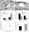
Ultrastructural analysis of synaptic boutons lacking postsynaptic β-Spectrin. (A) A cross section of a wild-type synaptic bouton. The postsynaptic muscle membrane folds (SSR) and the muscle fibers (m) are indicated. The presynaptic bouton is completely surrounded by SSR. A representative active zone is demarcated by arrows. (B) A cross section of a synaptic bouton from an animal lacking postsynaptic β-Spectrin. The SSR is severely reduced and almost absent above and below the presynaptic bouton. At many places, including active zones, muscle fibers directly abut the presynaptic membrane (asterisks). The boundaries of a representative active zone are indicated by arrows. (C) Analysis of SSR thickness in wild-type animals and in animals lacking postsynaptic β-Spectrin. (D) Analysis of SSR density in wild type and in animals lacking postsynaptic β-Spectrin. (E) Cumulative probability plot of active zone sizes in wild-type animals and in animals lacking postsynaptic β-Spectrin. (F) Quantification of active zone sizes (wild type = 531 ± 22 nm and β-spectrin = 828 ± 59 nm; P < 0.001). Error bars represent the SEM. Bars: (A and B) 500 nm.
Figure 4.
Figure 4.
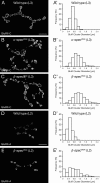
Analysis of postsynaptic glutamate receptor clusters at synapses lacking postsynaptic α- or β-Spectrin. (A–C and A′–C′) Analysis of postsynaptic glutamate receptor clusters in third instar larvae (L3). (A) A wild-type synapse on muscle 4, stained for the GluRII-C subunit of the postsynaptic glutamate receptor clusters. (A′) Frequency distribution of glutamate receptor cluster sizes in wild type. (B) Synapses lacking postsynaptic α-Spectrin show a severe perturbation of glutamate receptor clusters. (B′) The frequency distribution reveals a shift toward larger cluster sizes compared with wild type. (C) Synapses lacking postsynaptic β-Spectrin show increased glutamate receptor cluster size and altered organization. (C′) The analysis of the frequency distribution reveals a further increase in the number of larger cluster sizes compared with α-spectrin and wild type. (D–E′) Analysis of postsynaptic glutamate receptor clusters in second instar larvae (L2). (D) A wild-type synapse on muscle 4, stained for the GluRII-A subunit of the postsynaptic glutamate receptor clusters. (D′) Frequency distribution of glutamate receptor cluster sizes. (E) A second instar synapse lacking postsynaptic β-Spectrin shows an increase in glutamate receptor cluster size. (E′) The frequency distribution reveals a significant increase in cluster size compared with second instar wild-type animals. Bars, (A–E) 10 μm.
Figure 5.
Figure 5.
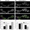
Postsynaptic Spectrin organizes pre- and postsynaptic synapse markers. (A) A wild-type synapse on muscle 4, stained for the presynaptic active zone marker nc82 and the postsynaptic glutamate receptor cluster subunit GluRII-C. (B) A synapse on muscle 4 lacking postsynaptic β-Spectrin. Multiple puncta of the presynaptic active zone marker nc82 accumulate opposite single, enlarged postsynaptic GluRII-C receptor clusters. (C) Quantification of presynaptic nc82 and postsynaptic GluRII-C puncta reveals a significant increase in the ratio of presynaptic nc82 puncta per postsynaptic glutamate receptor cluster in both L2 and L3 larvae in the absence of postsynaptic β-Spectrin, compared with wild type. (D) Quantification of presynaptic nc82 puncta and postsynaptic glutamate receptor clusters per NMJ area. Error bars represent the SEM. Bars, (A and B) 10 μm.
Figure 6.
Figure 6.
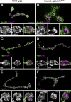
Postsynaptic Spectrin is required for the organization of active zone and periactive zone components. All images are of NMJs at muscle 4. Smaller images show higher magnifications and single channels from the areas indicated by the asterisks. (A) A wild-type synapse stained for the postsynaptic density marker GluRII-C and the postsynaptic periactive zone reporter Sh-GFP. Sh-GFP staining circumscribes the evenly distributed GluRII-C receptor clusters. (B) At a synapse lacking postsynaptic α-Spectrin, Sh-GFP is no longer confined to the area directly surrounding the postsynaptic receptor clusters and loses its latticelike appearance. (C) A wild-type synapse stained for the postsynaptic active zone marker Pak and the transsynaptic cell adhesion molecule Fas II, which localizes to the periactive zone. Fas II forms a honeycomb-like network that precisely surrounds Pak staining. (D) At synapses lacking postsynaptic β-Spectrin, Pak clusters become bigger and less regular shaped. The Fas II network is less regular, but still surrounds Pak-positive clusters. (E) A wild-type synapse stained for Fas II and the cytoplasmic presynaptic periactive zone marker Nwk. Fas II and Nwk partially colocalize and surround the presynaptic active zone. (F) At synapses lacking postsynaptic β-Spectrin, Fas II and Nwk lose their regular organization. Bars: (A–F) 10 μm; (magnifications) 3 μm.
Figure 7.
Figure 7.
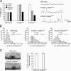
Synapses lacking postsynaptic α- or β-Spectrin show an increase in quantal size. (A) Quantification of the average mepsp amplitude (quantal size), EPSP amplitude, and quantal content in wild type and in animals lacking postsynaptic α- or β-Spectrin. Measurements are normalized to wild type to allow display of the data on a single graph. At synapses lacking postsynaptic α- or β-Spectrin there is a significant increase in quantal size compared with wild type (82% increase for α-spectrin and 85% increase for β-spectrin; P < 0.001). At synapses lacking postsynaptic α-Spectrin, EPSP amplitudes are larger than wild type, and there is no change in quantal content compared with wild type. At synapses lacking postsynaptic β-Spectrin, EPSPs are larger than wild type, but there is a significant decrease in quantal content. Representative mepsp traces are shown. (B) Histograms of mepsp amplitude distributions in wild type and in animals lacking postsynaptic α- or β-Spectrin. (C) Ultrastructural analysis of average synaptic vesicle diameters for wild-type synapses and synapses lacking postsynaptic β-Spectrin. There is no change in the average vesicle diameter at synapses lacking postsynaptic β-Spectrin compared with wild type. Representative images of active zones are shown at left. Error bars represent the SEM.
Figure 8.
Figure 8.
Postsynaptic α- and β-Spectrin are required for the normal localization of postsynaptic Ankyrin. (A–C) A wild-type synapse on muscle 4 stained for Ank and Dlg. Ank is present in the T-tubules throughout the muscles and highly enriched in the SSR of 1b and 1s boutons. (D–F) The postsynaptic expression of ank dsRNA results in the elimination of Ank in the muscle. Ank can no longer be detected in the T-tubules or the SSR. Presynaptic Ank becomes evident in the absence of postsynaptic Ank. Dlg organization within the postsynaptic SSR remains unaltered in the absence of postsynaptic Ank. (G–I) A muscle 4 synapse that lacks postsynaptic β-Spectrin. The SSR is clearly disturbed and Dlg organization is disrupted. Ank is almost completely absent from the SSR, but is still present in the T-tubule network (G). (J–L) A muscle 4 synapse that lacks postsynaptic α-Spectrin. The organization of the SSR is severely disturbed but Ank is still present at the SSR and colocalizes with Dlg. Bar, (A–L) 10 μm.
Figure 9.
Figure 9.
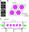
A postsynaptic Spectrin–actin network can define synapse dimensions and spacing. (A) Staining of GluRII-C receptors and Sh-GFP (Fig. 6). (B) Top view of a proposed α-/β-Spectrin hexagonal network. In this model, a unit of six Spectrin heterotetramers linked by actin filaments could participate in the organization of synapse size and spacing. (C) Side view of a single synapse. Spectrin heterotetramers are linked through interactions of β-Spectrin with short actin filaments. The postsynaptic Spectrin–actin network could participate in the localization of periactive zone proteins such as Dlg to the borders of the active zone. Dlg, in turn, could then bind and scaffold additional periactive zone proteins, including Fas II and Shaker.

Similar articles

Cited by

References

    1. Albin, S.D., and G.W. Davis. 2004. Coordinating structural and functional synapse development: postsynaptic p21-activated kinase independently specifies glutamate receptor abundance and postsynaptic morphology. J. Neurosci. 24:6871–6879. - PMC - PubMed
    1. Ango, F., G. di Cristo, H. Higashiyama, V. Bennett, P. Wu, and Z.J. Huang. 2004. Ankyrin-based subcellular gradient of neurofascin, an immunoglobulin family protein, directs GABAergic innervation at Purkinje axon initial segment. Cell. 119:257–272. - PubMed
    1. Atwood, H.L., C.K. Govind, and C.F. Wu. 1993. Differential ultrastructure of synaptic terminals on ventral longitudinal abdominal muscles in Drosophila larvae. J. Neurobiol. 24:1008–1024. - PubMed
    1. Bennett, V., and A.J. Baines. 2001. Spectrin and ankyrin-based pathways: metazoan inventions for integrating cells into tissues. Physiol. Rev. 81:1353–1392. - PubMed
    1. Bennett, V., J. Davis, and W.E. Fowler. 1982. Brain spectrin, a membrane-associated protein related in structure and function to erythrocyte spectrin. Nature. 299:126–131. - PubMed

Publication types

MeSH terms