. 2006 Nov 14;103(46):17266-71.
doi: 10.1073/pnas.0608156103. Epub 2006 Nov 6.

Self-renewal of embryonic stem cells by a small molecule

Affiliations

Self-renewal of embryonic stem cells by a small molecule

Shuibing Chen et al. Proc Natl Acad Sci U S A. .

Abstract

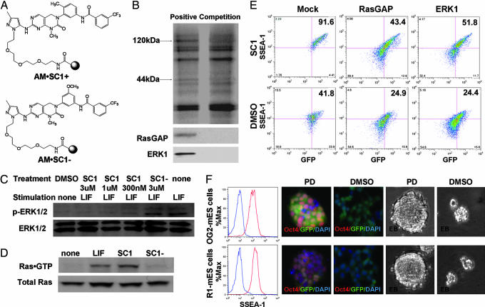
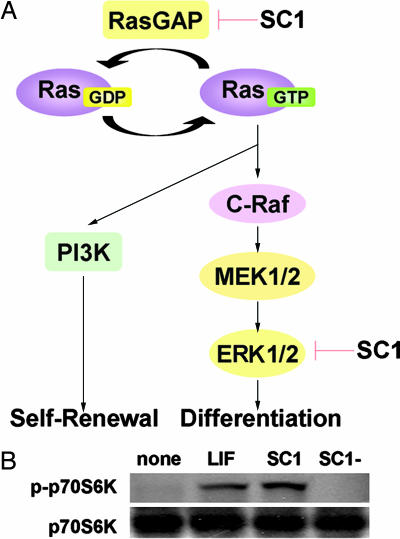
A cell-based screen of chemical libraries was carried out to identify small molecules that control the self-renewal of ES cells. A previously uncharacterized heterocycle, SC1, was discovered that allows one to propagate murine ES cells in an undifferentiated, pluripotent state under chemically defined conditions in the absence of feeder cells, serum, and leukemia inhibitory factor. Long-term SC1-expanded murine ES cells can be differentiated into cells of the three primary germ layers in vitro and also can generate chimeric mice and contribute to the germ line in vivo. Biochemical and cellular experiments suggest that SC1 works through dual inhibition of RasGAP and ERK1. Molecules of this kind may not only facilitate practical applications of stem cells in research and therapy, but also provide previously undescribed insights into the complex biology of stem cells.


Conflict of interest statement

The authors declare no conflict of interest.

Figures

Scheme 1.
Scheme 1.
Chemical structures of 3,4-dihydropyrimido[4,5-d]pyrimidine scaffold (A) and SC1 (B).
Fig. 1.
Fig. 1.
SC1-expanded mES cells maintain their ability to self-renew under feeder cell/LIF-free conditions. (A and B) Serially passaged, SC1-expanded OG2-mES cells maintain the expression of pluripotency markers by FACS analysis (A) (undifferentiated population: SC1 + SR, 95.9%; DMSO + SR, 1.2%; SC1 + N2B27, 95.7%; DMSO + N2B27, 0.6%; SC1 + N2, 95.8%; DMSO + N2, 0.1%; LIF + BMP4, 87.4%), histocytochemistry (alkaline phosphatase) and by RT-PCR (B) (Nanog, Oct4, and Sox2). mES cells were seeded in gelatin-coated six-well plates at 1.6 × 10⁴ cells/cm2 and maintained with 3 μM SC1 in the ESC-SR media, 1 μM SC1 in the ESC-N2B27 media, or 300 nM SC1 in the ESC-N2 media without LIF or feeder cells. mES cells (seeded at 1.6 × 10⁴ cells/cm2) maintained with 103 units/ml LIF plus 10 ng/ml BMP4 in the ESC-N2B27 media were used as a control. mES cells cultured with DMSO after two passages were used as negative control. Cells were split every 3 days and seeded at the same density (1.6 × 10⁴ cells/cm2 in six-well plates). SC1-expanded mES cells at passage 11 were analyzed with FACS, histocytochemistry, and RT-PCR. (C) Growth curves of OG2-mES cells under different culture conditions. mES cells maintained under the feeder condition were used as a positive control. Cells were split every 3 days, and cell numbers were counted by hemocytometer. Under all conditions, >95% cells maintain GFP expression by FACS analysis (Fig. 5D). (D and E) OG2-mES cells can be expanded by SC1 at low cell density (D) and maintain the expression of pluripotency markers after three passages (E) (undifferentiated population: ESC-SR, 90.0%; ESC-N2B27, 97.4%). OG2-mES cells were seeded at a low cell density (50 cells/cm2) and cultured with 3 μM SC1 in ESC-SR media or 1 μM SC1 in ESC-N2B27 media for three passages. Cells were split every 3 days and seeded at the same density (50 cells/cm2). Cell numbers at each passage were counted by hemocytometer. Cells at day 9 were analyzed with FACS. (F) SC1 (1 μM) inhibits differentiation of OG2-mES cells induced by 20% FBS (undifferentiated population: SC1, 89.5%; DMSO, 0.2%) or 1 μM RA (undifferentiated population: SC1, 93.4%; DMSO, 0.4%). OG2-mES cells were seeded at 1.6 × 10⁴ cells/cm2 and cultured with 20% FBS or 1 μM RA in the ESC-N2B27 media with/without 1 μM SC1 for 4 days (FBS) or 2 days (RA) and analyzed with FACS and histocytochemistry. (G) In vitro differentiation potential of SC1-expanded (1 μM concentration in ESC-N2B27 media), passage 11 OG2-mES cells. Neuronal, cardiac muscle, and endodermal differentiation were carried out by using established protocols and cells were stained with antibodies against β III-tubulin, myosin heavy chain, and Sox17, respectively (red, β III-tubulin, myosin heavy chain; green, Sox17; blue, DAPI) and analyzed by RT-PCR. Feeder-cultured OG2-mES cells were used as a control. (H) Germ-line contribution of SC1-expanded (3 μM concentration in ESC-SR media), passage 11 OG2-mES cells. Phase contrast and fluorescent images of female and male gonad (14.5 days postcoitum) and squeezed male gonad tissue clearly show the singular germ cells that express GFP driven by the Oct4 promoter (a marker for ES cells or embryonic germ cells). (SR, ESC-SR media; N2B27, ESC-N2B27 media; N2, ESC-N2 media; ALP, alkaline phosphatase; MHC, myosin heavy chain).
Fig. 2.
Fig. 2.
The effects of SC1 on known self-renewal pathways. (A) Western blot analysis of STAT3 phosphorylation at Tyr-705. OG2-mES cells were cultured in the ESC-N2B27 media overnight and then stimulated with DMSO (negative control), 1 μM SC1, 1 μM SC1− (negative control of SC1), 103 units/ml LIF (positive control), or 1 μM SC1 plus 103 units/ml LIF for 20 min. (B) Quantitative RT-PCR analysis of Id1 gene expression. OG2-mES cells were cultured in the ESC-N2B27 media overnight and then stimulated with DMSO (negative control), 1 μM SC1, or 50 ng/ml BMP4 (positive control) for 45 min. (C) Wnt signaling by Super (8×) TOPflash reporter assay. Stable Super (8×) TOPflash reporter 293T cells were treated with DMSO (negative control), 3 μM SC1, or 2 μM BIO (positive control) for 48 h and assayed for luciferase activity. (D) Transient activation of Nanog expression. OG2-mES cells were treated with 1 μM SC1 in the ESC-N2B27 media, and mRNAs were isolated at 0, 6, and 18 h, separately. In these experiments, OG2-mES cells were seeded at the same density (1.6 × 10⁴ cells/cm2). PD, PD098059; SC1−, negative control of SC1.
Fig. 3.
Fig. 3.
Affinity chromatography and target confirmation. (A) Structures of the positive affinity matrix (AM·SC1+) and the negative affinity matrix (AM·SC1−). (B) Silver staining and Western blot analysis of proteins retained by affinity supports. (C) SC1 inhibits autophosphorylation of ERK1/2 at Thr-202/Tyr-204. OG2-mES cells were cultured in the ESC-N2B27 media overnight and then stimulated with 103 units/ml LIF for 15 min in the presence or absence of SC1 or SC1−. (D) SC1 activates Ras activity. OG2-mES cells were treated with 1 μM SC1, 103 units/ml LIF, or 1 μM SC1− in the ESC-N2B27 media for 6 h, and whole-cell lysates were analyzed with the Ras Activation Assay Kit. (E) Overexpression of ERK1 and RasGAP. OG2-mES cells (seeded at 1.6 × 10⁴ cells/cm2) transiently transfected (transfection efficiency is ≈50%) with ERK1 or RasGAP were cultured in the ESC-N2B27 media with/without 1 μM SC1 for 3 days and analyzed with FACS (undifferentiated population: mock, 41.8 ± 1.9%; ERK1, 24.9 ± 2.5%; RasGAP, 24.4 ± 2.9%; Mock + SC1, 91.6 ± 3.6%; ERK1 + SC1, 43.4 ± 2.1%; RasGAP + SC1, 51.8 ± 2.0%). Empty vector was used as a negative control. (F) OG2-mES cells and R1-mES cells infected with lentivirus of shRasGAP were purified and cultured in the ESC-N2B27 media with 40 μM PD098059 for >10 passages. Stable cell lines of GFPhigh population were seeded at 1.6 × 10⁴ cells/cm2 and cultured with/without PD098059 (40 μM) in the ESC-N2B27 media without feeder cells, LIF, and serum. Cells were split every 3 days. Cells with PD098059 treatment at passage 11, and cells without PD098059 treatment at passage 6 were analyzed with FACS (red line, with PD098059; blue line, without PD098059), immunocytochemistry (red, Oct4; green, GFP; blue, DAPI), and EB formation.
Fig. 4.
Fig. 4.
Mechanism of action. (A) Proposed mechanism of action of SC1. (B) SC1 activates p70S6K phosphorylation. OG2-mES cells were treated with 103 units/ml LIF, 1 μM SC1, or 1 μM SC1− in the ESC-N2B27 media for 10 h.

References

    1. Ding S, Wu TY, Brinker A, Peters EC, Hur W, Gray NS, Schultz PG. Proc Natl Acad Sci USA. 2003;100:7632–7637. - PMC - PubMed
    1. Niwa H, Burdon T, Chambers I, Smith A. Genes Dev. 1998;12:2048–2060. - PMC - PubMed
    1. Ying QL, Nichols J, Chambers I, Smith A. Cell. 2003;115:281–292. - PubMed
    1. Qi X, Li TG, Hao J, Hu J, Wang J, Simmons H, Miura S, Mishina Y, Zhao GQ. Proc Natl Acad Sci USA. 2004;101:6027–6032. - PMC - PubMed
    1. Avilion AA, Nicolis SK, Pevny LH, Perez L, Vivian N, Lovell-Badge R. Genes Dev. 2003;17:126–140. - PMC - PubMed

Publication types