. 2006 Nov 14;103(46):17432-7.
doi: 10.1073/pnas.0607939103. Epub 2006 Nov 6.

An essential role of NF-kappaB in the "tumor-like" phenotype of arthritic synoviocytes

Affiliations

An essential role of NF-kappaB in the "tumor-like" phenotype of arthritic synoviocytes

Xiangli Li et al. Proc Natl Acad Sci U S A. .

Abstract

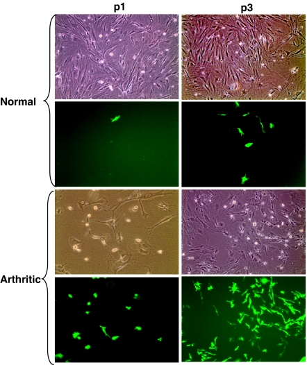
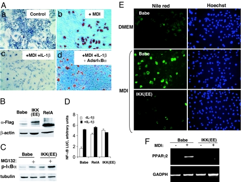
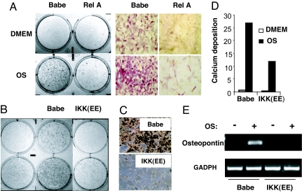
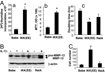
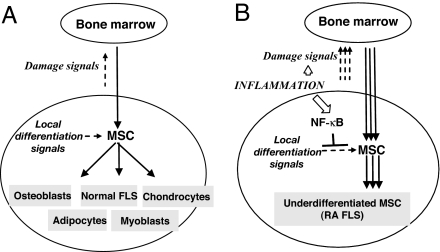
A hallmark of rheumatoid arthritis is the formation of an aggressive, tumor-like structure called pannus that erodes the joint. A major cellular component of the pannus is the fibroblast-like synoviocyte (FLS), whose morphology strikingly resembles that of a transformed cell, but underlying mechanisms of this "transformation" are not known. Here, using animal models of rheumatoid arthritis, we show that arthritic FLS contain a substantial (>30%) fraction of bone marrow-derived precursors that can differentiate in vitro into various mesenchymal cell types, but inflammation prevents the multilineage differentiation. We show that the transcription factor NF-kappaB plays the key role in the repression of osteogenic and adipogenic differentiation of arthritic FLS. Furthermore, we show that specific activation of NF-kappaB profoundly enhances proliferation, motility, and matrix-degrading activity of FLS. We thus propose that arthritic FLS are mesenchymal stem cells whose differentiation is arrested at early stages of differentiation by activation of NF-kappaB.


Conflict of interest statement

The authors declare no conflict of interest.

Figures

Fig. 1.
Fig. 1.
The presence of BM-derived cells in the populations of normal and arthritic FLS. BM cells from GFP transgenic donor mice were transferred into lethally irradiated GFP-negative recipient mice. Two months later, primary FLS were established by an enzymatic dispersal of synovial explants of the recipient BMT mice. p1 and p3, the first and third passages of primary FLS. The first and third rows represent phase-contrast microscopy; the second and fourth rows represent fluorescent microscopy. Normal, primary FLS that were obtained from normal joints of the BMT mice. At the third passage, GFP-positive cells comprised 1.2 ± 0.2% of the population of normal FLS (mean ± SEM of two experiments). Arthritic, primary FLS that were obtained from arthritic BMT recipient mice as described in Materials and Methods. At the third passage, GFP-positive cells comprised 33.7 ± 1.6% of the population of arthritic FLS (mean ± SEM of three experiments). (Original magnification: ×100.)
Fig. 2.
Fig. 2.
NF-κB activation is necessary and sufficient to inhibit the adipogenic differentation of arthritic rFLS. (A) NF-κB is required for the antidifferentiating activity of IL-1 in the adipogenic differentiation of arthritic rFLS. Primary FLS from rats with PG-PS-induced arthritis were infected with a control, “empty” adenovirus (Control) (ac) or with adenovirus expressing superrepressor IκBα (AdsrIκBα) (d). Cells were incubated in DMEM (a) or in an adipogenesis-inducing medium (MDI) (b), or in the MDI medium in the presence of IL-1β (100 units/ml) (c and d). After 21 days, differentiated cells were visualized by staining the accumulated lipids with an Oil red stain. Representative data of three experiments are shown. (Original magnification: ×100.) (BD) Constitutive activation of NF-κB by retrovirus-mediated gene transfer of a dominant-positive IKKβ and RelA. Primary rFLS were transduced with a control RV Babe (control Babe) or with vectors that expressed cDNA of FLAG-tagged constitutively active IKKβ mutant [IKK(EE)], or FLAG-tagged RelA cDNA. After puromycin selection, NF-κB activation was assessed. (B) The expression of exogenous IKK(EE) and RelA proteins was assessed by immunoblotting with anti-FLAG Ab. (C) The expression of IKK(EE) results in accumulation of a phosphorylated form of IκBα. The control (Babe) and IKK(EE) cells were detected by using antiphospho IκBα Ab. Where indicated, cells were pretreated with a proteasomal inhibitor, MG132, to allow for accumulation of the phosphorylated IκBα. (D) Ectopic expression of IKK(EE) and RelA induces constitutive activation of NF-κB-driven gene expression. Transduced rFLS were transiently cotransfected with an NF-κB-inducible reporter vector expressing the firefly luciferase and a constitutively active reporter expressing the Renilla luciferase. Normalized values of NF-κB activation from duplicated samples are shown. Representative data of two experiments are shown. (E and F) Constitutive activation of NF-κB inhibits the adipogenic differentiation of arthritic rFLS. (E) RV-transduced cells (as in B) were incubated in a control medium (DMEM) or in an adipogenesis-inducing medium (MDI). After 21 days, the adipogenic differentiation was assessed by staining accumulated lipids with Nile red. The staining was visualized by fluorescent microscopy (Left). (Original magnification: ×100.) Cells were counterstained with the nuclear stain Hoechst 33342 (Right). (F) The expression of an adipogenic differentiation marker (PPARγ2) was assessed in cells shown in B by RT-PCR.
Fig. 3.
Fig. 3.
Constitutive activation of NF-κB inhibits osteogenic differentiation of arthritic rFLS. RV-transduced cells (as in Fig. 2B) were incubated in a control medium (DMEM) or in an osteogenesis-inducing medium (OS). (A) The activity of ALP in the control (Babe) and in RelA-transduced cells after 14 days of incubation. (B) ALP activity in the control and IKK(EE)-transduced cells after 14 days of incubation. (C) Mineral deposition in the control (Babe) and in IKK(EE)-transduced cells after 21 days of incubation, as assessed by silver staining. (Original magnification in A and C: ×100.) (D) Calcium deposition in cells shown in C was quantitatively assessed and normalized on protein content. (E) The expression of an osteogenic differentiation marker (osteopontin) in cells shown in B as assessed by RT-PCR.
Fig. 4.
Fig. 4.
Constitutive activation of NF-κB accelerates cell growth and enhances the invasiveness of arthritic rFLS. (A) Constitutive activation of NF-κB accelerates cell growth. Cells were transduced as described for Fig. 2B, and proliferation was assessed by determining thymidine incorporation (a) or by MTT proliferation assay (b). (Ac) To directly assess cell growth, cells were plated at a low density and counted after 5 days in culture. The numbers of cells was adjusted to plating efficacy that was assessed 1 day after plating. Doubling times were calculated assuming an exponential cell growth. Representative data of three experiments are shown. ∗, P < 0.05 vs. Babe control. (B) The production of preproactive and active forms of MMP-13 protein by transduced cells was assessed by immunoblotting in lysates of cells that were unstimulated or stimulated for 4 h with IL-1β (100 units/ml). The blot was reprobed with β-actin Ab. (C) Constitutive activation of NF-κB enhances invasiveness. Cell motility was assessed in a transwell cell migration assay as described in Materials and Methods. The cells that migrated through the filter were stained and counted. One representative experiment from three independent experiments is shown.
Fig. 5.
Fig. 5.
Constitutive activation of NF-κB in arthritic joints maintains the highly invasive, undifferentiated phenotype of arthritic FLS.

References

    1. Firestein GS. Nature. 2003;423:356–361. - PubMed
    1. Pap T, Muller-Ladner U, Gay RE, Gay S. Arthritis Res. 2000;2:361–367. - PMC - PubMed
    1. Edwards JC. Arthritis Res. 2000;2:344–347. - PMC - PubMed
    1. Firestein GS. Arthritis Rheum. 1996;39:1781–1790. - PubMed
    1. Muller-Ladner U, Kriegsmann J, Gay RE, Gay S. Rheum Dis Clin North Am. 1995;21:675–690. - PubMed

Publication types

MeSH terms

LinkOut - more resources