. 2006 Nov 14;103(46):17450-5.
doi: 10.1073/pnas.0604026103. Epub 2006 Nov 6.

Angiopoietin-like protein 4 converts lipoprotein lipase to inactive monomers and modulates lipase activity in adipose tissue

Affiliations

Angiopoietin-like protein 4 converts lipoprotein lipase to inactive monomers and modulates lipase activity in adipose tissue

Valentina Sukonina et al. Proc Natl Acad Sci U S A. .

Abstract

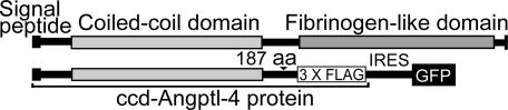
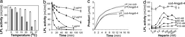
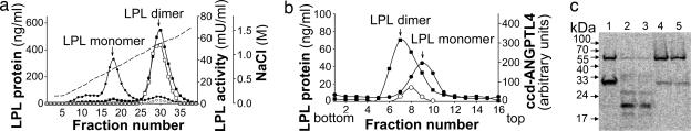
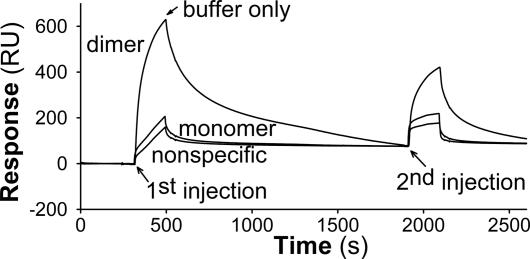
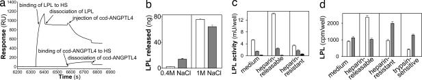
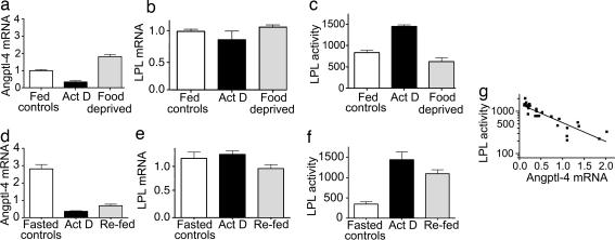
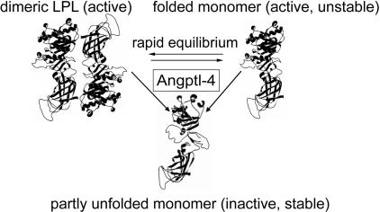
Lipoprotein lipase (LPL) has a central role in lipoprotein metabolism to maintain normal lipoprotein levels in blood and, through tissue specific regulation of its activity, to determine when and in what tissues triglycerides are unloaded. Recent data indicate that angiopoietin-like protein (Angptl)-4 inhibits LPL and retards lipoprotein catabolism. We demonstrate here that the N-terminal coiled-coil domain of Angptl-4 binds transiently to LPL and that the interaction results in conversion of the enzyme from catalytically active dimers to inactive, but still folded, monomers with decreased affinity for heparin. Inactivation occurred with less than equimolar ratios of Angptl-4 to LPL, was strongly temperature-dependent, and did not consume the Angptl-4. Furthermore, we show that Angptl-4 mRNA in rat adipose tissue turns over rapidly and that changes in the Angptl-4 mRNA abundance are inversely correlated to LPL activity, both during the fed-to-fasted and fasted-to-fed transitions. We conclude that Angptl-4 is a fasting-induced controller of LPL in adipose tissue, acting extracellularly on the native conformation in an unusual fashion, like an unfolding molecular chaperone.


Conflict of interest statement

The authors declare no conflict of interest.

Figures

Fig. 1.
Fig. 1.
A schematic view of the plasmid used to express the coiled-coil domain of murine Angptl-4. (Upper) Overview of the structure of angiopoietin-like proteins. (Lower) The pIRES-hrGFPII expression vector containing the coding sequences for the coiled-coil domain of Angptl-4 (ccd-Angptl-4, residues 1–187) fused at the 3′ end with a 3× FLAG-tag sequence.
Fig. 2.
Fig. 2.
Inhibition of LPL by purified ccd-Angptl-4 depended on temperature and concentration, but the ccd-Angptl-4 was not consumed, and the inhibition was not relieved by heparin. (a) LPL (350 ng/ml) was incubated at the indicated temperatures with (filled bars) or without (open bars) ccd-Angptl-4 (1 μg/ml) in 20 mM Tris·Cl/0.15 M NaCl, pH 7.4, in the presence of 10% FCS. After 1 h, the remaining LPL activity was determined. (b) LPL was incubated without or with the indicated concentrations of ccd-Angptl-4 at 25°C under the same conditions as in a. Samples from the mixtures were analyzed for remaining LPL activity at the indicated times. (c) Inactivation was studied in real time by following hydrolysis of triacetin (in 0.15 M NaCl, pH 7.4) by LPL (6 μg/ml). The released acetic acid was titrated with NaOH by using a pH-stat. The spontaneous hydrolysis of triacetin (without addition of LPL) was subtracted. Upper curves show LPL without ccd-Angptl-4; lower curves show LPL in the presence of ccd-Angptl-4. The molar ratio of ccd-Angptl-4 (calculated from the monomer molecular mass) to LPL dimers was ≈1:1 for the first recording (1st). After LPL had been fully inactivated, a second addition of the same amount of active LPL was added to the reactions (2nd), and the progress of hydrolysis was again followed with time. A third (3rd) addition of LPL was made to the reaction vessel with ccd-Angptl-4. (d) LPL (4 nM) was incubated without or with the indicated concentrations of ccd-Angptl-4 at 25°C under the same conditions as in a, in the presence of the indicated concentrations of heparin.
Fig. 3.
Fig. 3.
Inhibition of LPL by ccd-Angptl-4 involved conversion of active, dimeric LPL to inactive LPL monomers. (a) Partially inactivated LPL was separated by chromatography on heparin-Sepharose. For this experiment, a mixture of 200 ng of 125I-labeled and 1.8 μg of unlabeled LPL was incubated with 2.4 μg of ccd-Angptl-4 (circles) or buffer (20 mM Tris·Cl/0.15 M NaCl) (squares) for 30 min at 20°C in the presence of 10% delipidated FCS. The mixtures were then applied on a 1-ml Hi-Trap heparin-Sepharose column and eluted by a gradient of NaCl (dashed line). Fractions of 0.5 ml were collected, and LPL radioactivity (filled symbols) and LPL activity (open symbols) was measured. The positions of the peaks for a standard mixture of dimeric and monomeric LPL are indicated by arrows. ccd-Angptl-4, run in the absence of LPL and detected by Western blots with anti-FLAG antibodies, eluted at ≈0.4 M NaCl (data not shown). (b) Partially inactivated LPL was separated by sucrose-density gradient ultracentrifugation. For this experiment, 17.5 ng of 125I-labeled LPL and 900 ng of unlabeled LPL in 10% delipidated FCS were incubated for 30 min at 20°C with 2.5 μg of ccd-Angptl-4 (filled circles) or buffer (20 mM Tris·Cl/0.15 M NaCl) (filled squares) in a total volume of 0.5 ml. Then 0.2 ml of each sample was applied to linear sucrose-density gradients. The curves represent mean values from two tubes run in parallel for each sample. The profile for sedimentation of ccd-Angptl-4 was determined by scanning of a Western blot of samples from the fractions visualized by an anti-FLAG antibody (open circles). (c) The conformation of partially inactivated LPL was probed by limited tryptic digestion. For this experiment, samples of 125I-labeled LPL dimers and monomers isolated by heparin-Sepharose chromatography after thermal inactivation at 37°C or inactivation by treatment with ccd-Angptl-4 (as in a) were incubated for 15 min with trypsin and were then analyzed by SDS/PAGE under reducing conditions. The bands were detected by imaging. Lane 1 is a reference sample of active dimeric LPL treated with trypsin. Lane 2 is the peak of inactive LPL (first peak in a) treated with trypsin. Lane 4 is the peak of active dimeric LPL (second peak in a) treated with trypsin. Lanes 3 and 5 are analogous to lanes 2 and 4, but, in this case, LPL was partially inactivated by thermal treatment before separation on heparin-Sepharose and trypsin treatment.
Fig. 4.
Fig. 4.
ccd-Angptl-4 bound with high affinity to dimeric, but less to monomeric, LPL as determined by surface plasmon resonance. Biotinylated dimeric and monomeric LPL were bound to streptavidin-coated sensor chips to a level of 5,100 response units (RU) and 5,300 RU, respectively. ccd-Angptl-4 (13.8 μg/ml) was injected over these two flow cells and over a flow cell that contained only streptavidin (1st injection). At the indicated time (arrow) the flow was changed to buffer without ccd-Angptl-4 to study dissociation of the ligand. Then a second injection of the same ccd-Angptl-4 solution was made (2nd injection). The buffer contained 20 mM Hepes and 0.15 M NaCl and the temperature was 25°C.
Fig. 5.
Fig. 5.
Binding of LPL to immobilized HS/heparin or to cell surfaces did not protect the enzyme from inactivation by ccd-Angptl-4. (a) Biotinylated HS was immobilized to two flow cells of a streptavidin-coated sensor chip. Dimeric LPL (50 μg/ml) was bound to one of these flow cells (upper curve). ccd-Angptl-4 (17 μg/ml) was simultaneously injected over the flow cell with already bound LPL and the flow cell with only HS (lower curve). The times for the injections and for the wash with running buffer (dissociation phase) are shown by arrows. (b) Heparin-Sepharose beads, preloaded with 125I-labeled LPL, were incubated with conditioned medium from 293T cells transfected with pIRES-hrGFPII vector (open bars) or pIRES-ccd-Angptl-4 (filled bars). The beads were then sequentially eluted with buffer containing 0.4 and 1 M NaCl. The amount of LPL released in each fraction was determined from the radioactivity that was recalculated to LPL mass (ng). The data are means of five parallel samples. (c and d) LPL activity (c) and LPL mass (d) in fractions obtained from 3T3-L1 preadipocytes loaded with 125I-labeled LPL and then incubated with (filled bars) or without (open bars) ccd-Angptl-4 for 1 h at 25°C. The results represent mean values of 6 wells for activity and 18 wells for radioactivity ± SD. One thousand cpm corresponded to 6.8 ng of LPL. Activity of endogenously produced LPL in cells incubated without [125I]LPL was also measured (filled bars). Surface exposure of the heparin-resistant radioactivity remaining with the cells was probed by brief digestion with trypsin and measurement of release into the medium. Less than 47 ± 8.7 cpm and 68 ± 14 cpm, respectively, was released by the buffer without trypsin.
Fig. 6.
Fig. 6.
The levels of Angptl-4 mRNA in adipose tissue changed rapidly with nutritional state. (a and d) Angptl-4 mRNA abundance. (b and e) LPL mRNA abundance. (c and f) LPL activity. (ac) The fed-to-fasted transition. For this experiment, food was removed from two groups of rats in the morning. The rats in one of these groups were given an i.p. injection of Actinomycin D (2 mg/kg of body weight). The rats were killed 6 h later. (df) The same parameters for the fasted-to-fed transition. For this experiment, food was removed from the rats at 4 p.m. the day before the experiment. The next morning, food was returned to two groups of rats. The rats in one of these groups were given an i.p. injection of Actinomycin D. The rats were killed 6 h later. There were three to six rats in each group. Data for LPL activity are shown as mean ± SEM. Data for expression of Angptl-4 and LPL mRNA are shown as mean ± SD. (g) The relation between Angptl-4 mRNA abundance and LPL activity for all individual rats. Note that the y axis is logarithmic. Correlation analysis for the log of LPL activity vs. Angptl mRNA abundance returned P < 0.0001, Pearson r = −0.88.
Fig. 7.
Fig. 7.
Schematic illustration of possible mechanisms for action of Angptl-4 on LPL. The active form of LPL is a noncovalent dimer with propensity to dissociate to inactive, but still folded, monomers. Kinetic analyses have shown that inactivation most likely involves a short-lived intermediate active monomer (21, 23). It is possible that Angptl-4 interacts preferentially with this intermediate state and thereby drives LPL from the active dimer to inactive monomers. It is also possible that Angptl-4 interacts with the active LPL dimers and promotes their dissociation to inactive monomers or dissociation to the active intermediates, which, in turn, refold to inactive monomers. The schematic representation of the LPL subunit is based on the model generated by van Tilbeurgh et al. (24).

References

    1. Olivecrona T, Olivecrona G. In: Lipoproteins in Health and Disease. Betteridge DJ, Illingworth DR, Shepherd J, editors. London: Arnold; 1999. pp. 223–246.
    1. Preiss-Landl K, Zimmermann R, Hammerle G, Zechner R. Curr Opin Lipidol. 2002;13:471–481. - PubMed
    1. Merkel M, Eckel RH, Goldberg IJ. J Lipid Res. 2002;43:1997–2006. - PubMed
    1. Osborne JC, Jr, Bengtsson-Olivecrona G, Lee NS, Olivecrona T. Biochemistry. 1985;24:5606–5611. - PubMed
    1. Enerbäck S, Semb H, Tavernier J, Bjursell G, Olivecrona T. Gene. 1988;64:97–106. - PubMed

Publication types

MeSH terms