. 2006 Dec;149(8):988-97.
doi: 10.1038/sj.bjp.0706953. Epub 2006 Nov 6.

Lovastatin protects human endothelial cells from the genotoxic and cytotoxic effects of the anticancer drugs doxorubicin and etoposide

Affiliations

Lovastatin protects human endothelial cells from the genotoxic and cytotoxic effects of the anticancer drugs doxorubicin and etoposide

J Damrot et al. Br J Pharmacol. 2006 Dec.

Abstract

Background and purpose: 3-Hydroxy-3-methylglutaryl-coenzyme A (HMG-CoA) reductase inhibitors (statins) are frequently used lipid-lowering drugs. Moreover, they exert pleiotropic effects on cellular stress responses and death. Here, we analysed whether lovastatin affects the sensitivity of primary human endothelial cells (HUVEC) to the anticancer drug doxorubicin.

Experimental approach: We investigated whether pretreatment of HUVEC with low dose of lovastatin influences the cellular sensitivity to doxorubicin. To this end, cell viability, proliferation and apoptosis as well as DNA damage-triggered stress response were analysed.

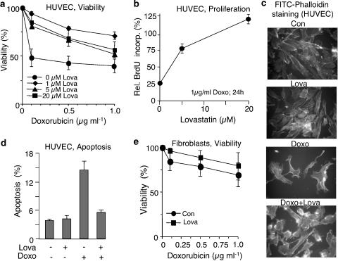
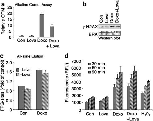
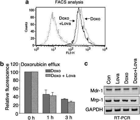
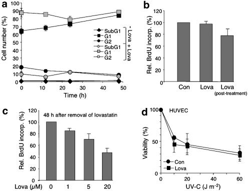
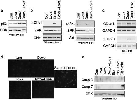
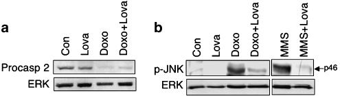
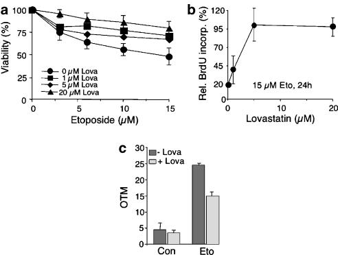
Key results: Lovastatin reduced the cytotoxic potency of doxorubicin in HUVEC. Lovastatin attenuated the doxorubicin-induced increase in p53 as well as activation of checkpoint kinase (Chk-1) and stress-activated protein kinase/c-Jun-N-terminal kinase (SAPK/JNK). Acquired doxorubicin resistance was independent of alterations in doxorubicin efflux and cell cycle progression. Also, doxorubicin-triggered production of reactive oxygen species (ROS) and formation of oxidative DNA lesions remained unaffected by lovastatin. However, lovastatin impaired DNA strand break formation induced by doxorubicin. Notably, lovastatin also conferred cross-resistance to the cytotoxic and genotoxic effects of etoposide, indicating that lovastatin shields topoisomerase II against poisons.

Conclusions and implications: Based on these data, we suggest that lovastatin-mediated resistance to topoisomerase II inhibitors is due to a reduction in DNA damage and, hence, it attenuates stress responses leading to cell death that are triggered by DNA damage. Therefore, lovastatin might be useful clinically for alleviating side-effects of anticancer therapies that include topoisomerase II inhibitors.


Figures

Figure 1
Figure 1
Lovastatin protects primary human endothelial cells from doxorubicin-induced cytotoxicity. (a) Primary human endothelial cells (HUVEC) were pretreated overnight with the indicated concentration of lovastatin. Afterwards, cells were exposed to different doses of doxorubicin. After incubation period of 48 h in the absence of lovastatin, cell viability was assayed using the WST assay as described in Methods. Data shown are the mean, and vertical lines show s.d., from three independent experiments each performed in triplicate. (b) HUVEC were pretreated overnight with different concentration of lovastatin. Afterwards, cells were treated with doxorubicin (1 μg ml−1). After incubation period of 24 h in the absence of lovastatin, cells were pulse-labelled with BrdU for 2 h. Incorporation of BrdU was assayed as described in Methods. BrdU labelling of doxorubicin-treated cells was related to that of the corresponding untreated control which was set to 100%. Data shown are the mean and s.d. from a representative experiment performed in quadruplicate. (c) HUVEC were pretreated overnight with lovastatin (1 μM) before they were exposed to doxorubicin (5 μg ml−1, 1 h treatment). After an incubation period of 48 h, morphological alterations of the actin cytoskeleton were analysed by FITC phalloidin staining followed by microscopic analysis. (d) After overnight pretreatment of HUVEC with lovastatin (Lova, 20 μM), doxorubicin (1 μg ml−1) was added and cells were post-incubated for 72 h in the absence of lovastatin, before the frequency of apoptotic death was determined by FACS-based annexin V method. Data shown are the mean and s.d. from three independent experiments. (e) Primary human fibroblasts were pretreated overnight with lovastatin (20 μM). Doxorubicin exposure and determination of cell viability were performed as described in (a).
Figure 2
Figure 2
Lovastatin lowers doxorubicin-induced genotoxicity in HUVEC. (a) After overnight pretreatment with lovastatin (1 μM), HUVEC were exposed to doxorubicin (5 μg ml−1). After an incubation period of 1 h, cells were harvested and DNA strand break formation was investigated by the comet assay as described in Methods. Shown are the mean and s.d. (vertical lines) from at least two independent experiments. (b) HUVEC were pretreated with lovastatin as described in (a). One hour after doxorubicin exposure (5 μg ml−1), phosphorylated histone (γ-H2AX) was determined in nuclear extracts. As a loading control, the filter was reprobed with ERK2-specific antibody (ERK). (c) Oxidative DNA damage was assayed by measuring FPG-sensitive sites in the genomic DNA. After lovastatin pretreatment (1 μM, overnight) cells were exposed to doxorubicin (10 μg ml−1) and the level of oxidative DNA damage was quantified one hour later as described in Methods. Data shown are the mean and s.d. (vertical lines) from three independent experiments each performed in duplicate. (d) Cells were left untreated (Con) or were pretreated overnight with lovastatin (Lova; 1 μM) before doxorubicin was added (5 μg ml−1). ROS formation was quantitated upon addition of DCF in 30 min time intervals (up to 90 min) as described in Methods. As positive control, cells were treated for 1 h with hydrogen peroxide (H2O2, 0.01%). Data shown are the mean and s.d. from a representative experiment performed in quadruplicate.
Figure 3
Figure 3
Lovastatin mediated protection is independent from uptake and efflux of doxorubicin. (a) HUVEC were left untreated or were pretreated overnight with lovastatin (1 μM) before addition of doxorubicin (5 μg ml−1). One hour after treatment, doxorubicin uptake was determined by FACS-based analysis of doxorubicin fluorescence. Shown is the result of one representative experiment out of three. (b) Lovastatin pretreated (1 μM, overnight)- or untreated HUVEC were exposed to doxorubicin as described in (a). Afterwards, doxorubicin was removed and doxorubicin fluorescence was determined 1 and 3 h later by FACS. Data shown are the mean and s.d. (vertical lines) from at least two independent experiments each performed in duplicate. (c) Analysis of mRNA expression of two main drug transporters, namely mdr-1 and mrp-1. GAPDH mRNA expression was analysed as an internal control.
Figure 4
Figure 4
Effect of lovastatin on cell cycle progression of HUVEC. (a) HUVEC were pretreated overnight with lovastatin (1 μM)(+Lova) or were left untreated (−Lova). Afterwards, fresh medium was added. After a further incubation period of up to 48 h, cells were harvested and cell cycle distribution was analysed by FACS. Data shown are the mean and s.d. from at least two independent experiments. (b) After overnight pretreatment of HUVEC with lovastatin (1 μM) fresh medium containing BrdU was added and cells were further incubated for 3 h in the absence (Lova) or presence (Lova, post-treatment) of lovastatin. BrdU incorporation was quantified by ELISA as described in Methods. Data shown are mean and s.d. from one representative experiment performed in quadruplicate. Con, untreated cells. (c) HUVEC were pretreated overnight with different concentration of lovastatin as indicated. Afterwards, medium was replaced by fresh medium. After a postincubation period of 48 h, cells were pulse-labelled with BrdU for 2 h. BrdU incorporation was quantified by ELISA as described in Methods. Data show are the mean and s.d. from at least two independent experiments each performed in triplicate. (d) HUVEC were left untreated (Con) or were pretreated overnight with lovastatin (Lova). Afterwards, cells were irradiated with different doses of UV-C light. After incubation period of 48 h, cell viability was determined as described in Methods. Data shown are the mean and s.d. from at least two independent experiments each performed in triplicate. Under identical experimental conditions, lovastatin pretreatment rendered human fibroblasts more sensitive to UV-C irradiation (data not shown).
Figure 5
Figure 5
Lovastatin has pleiotropic inhibitory effects on doxorubicin-induced stress responses of HUVEC. (a, b) Lovastatin pretreated (1 μM, overnight) or untreated cells (Con) were exposed to doxorubicin (2 μg ml−1; 1 h pulse treatment). After a post-incubation period of 8 h, expression of p53 (a) and of phosphorylated (activated) forms of p-Chk-1 (phosphorylated at Ser345) and Akt kinase (p-Akt) (phosphorylated at Ser473) (b) were assayed by Western blot analysis. Shown are the autoradiograms. Expression levels of EKR2 (ERK) and non-phosphorylated Chk-1 and Akt, respectively, are included as loading controls. (c) Cells were treated with lovastatin and doxorubicin, as described above; 12 h after exposure, mRNA expression of Fas ligand (CD95L) and Fas receptor (CD95R) was analysed by RT–PCR. mRNA expression of GAPDH was used as an internal control. Shown are the ethidium bromide stained gels (inverse presentation). (d) HUVEC were pretreated or not (Con) with lovastatin (1 μM, overnight) before doxorubicin was added (5 μg ml−1, 1 h). After a post-incubation period of 48 h, activation of caspase-3 was analysed by immunohistochemistry as described in Methods. As a positive control, cells were treated with staurosporine (1 μM, 6 h). (e) HUVEC were pretreated with lovastatin and exposed to doxorubicin (5 μg ml−1, 1 h) or etoposide (12 μM, 1 h). Cisplatin (50 μM, 3 h) was used as a positve control. Activated forms of caspase-3 and -7 were analysed 72 h after genotoxin exposure by Western blot analysis. As a loading control, the filter was reprobed with anti-ERK2 (ERK) antibody. The autoradiogram is shown.
Figure 6
Figure 6
Effect of lovastatin on doxorubicin-triggered activation of caspase-2 and stress kinase (SAPK/JNK). (a) HUVEC, pretreated or not (Con) overnight with lovastatin (1 μM), were exposed to doxorubicin (5 μg ml−1, 1 h). After a post-incubation period of 72 h, expression levels of procaspase-2 were determined by Western blot analysis. Expression of ERK2 (ERK) protein was determined as a loading control. (b) HUVEC were pretreated with lovastatin (1 μM; overnight) and subsequently exposed to doxorubicin (2.5 μg ml−1). Six hours after doxorubicin exposure, activation of the stress kinase p46/JNK1 (p-JNK) (dual phosphorylation at Thr183/Tyr185) was determined by Western blot analysis using phosphospecific JNK antibody. The alkylating agent MMS (1 mM, 2 h) was used as a positive control (Fritz and Kaina (2006)).
Figure 7
Figure 7
Lovastatin confers cross-resistance to the topoisomerase II-specific inhibitor etoposide. (a) HUVEC were pretreated overnight with the indicated concentration of lovastatin before etoposide was added. After a post-incubation period of 48 h in the absence of the statin, cell viability was assessed using the WST assay, as described in Methods. Data shown are the mean and s.d. (vertical lines) from three independent experiments each performed in triplicate. (b) HUVEC were pretreated with different concentration of lovastatin (1–20 μM, overnight). Afterwards, cells were exposed to etoposide (15 μM). After an incubation period of 24 h (in the absence of lovastatin), cells were pulse-labelled with BrdU for 2 h. Incorporation of BrdU was assayed as described in Methods. BrdU labelling of irradiated cells was related to that of the corresponding non-irradiated control, which was set to at 100%. Data shown are the mean and s.d. from one representative experiment performed in quadruplicate. (c) HUVEC were left untreated (−Lova) or were pretreated overnight with 1 μM of lovastatin (+Lova) before addition of etoposide (15 μM). After a further incubation period of 1 h, cells were harvested and the level of DNA strand breaks was quantified by the comet assay, as described in Methods. Shown are the mean and s.d. from two independent experiments. Con, untreated cells.

References

    1. Adamson P, Marshall CJ, Hall A, Tilbrook PA. Post-translational modifications of p21rho proteins. J Biol Chem. 1992;267:20033–20038. - PubMed
    1. Bachur NR, Yu F, Johnson R, Hickey R, Wu Y, Malkas L. Helicase inhibition by anthracycline anticancer agents. Mol Pharmacol. 1992;41:993–998. - PubMed
    1. Brown GA, McPherson JP, Gu L, Hedley DW, Toso R, Deuchars KL, et al. Relationship of DNA topoisomerase II alpha and beta expression to cytotoxicity of antineoplastic agents in human acute lymphoblastic leukemia cell lines. Cancer Res. 1995;55:78–82. - PubMed
    1. Cafforio P, Dammacco F, Gernone A, Silvestris F. Statins activate the mitochondrial pathway of apoptosis in human lymphoblasts and myeloma cells. Carcinogenesis. 2005;26:883–891. - PubMed
    1. Canman CE, Kastan MB. Signal transduction. Three paths to stress relief. Nature. 1996;384:213–214. - PubMed

Publication types

MeSH terms