. 2006 Dec;149(8):1039-48.
doi: 10.1038/sj.bjp.0706900. Epub 2006 Nov 6.

No proarrhythmic properties of the antibiotics Moxifloxacin or Azithromycin in anaesthetized dogs with chronic-AV block

Affiliations

No proarrhythmic properties of the antibiotics Moxifloxacin or Azithromycin in anaesthetized dogs with chronic-AV block

M B Thomsen et al. Br J Pharmacol. 2006 Dec.

Abstract

Background & purpose: The therapeutically available quinolone antibiotic moxifloxacin has been used as a positive control for prolonging the QT interval in both clinical and non-clinical studies designed to assess the potential of new drugs to delay cardiac repolarization. Despite moxifloxacin prolonging QT, it has not been shown to cause torsades de pointes arrhythmias (TdP). Azithromycin is a macrolide antibiotic that has rarely been associated, clinically, with cases of proarrhythmia. As there is a lack of clinical data available, the cardiac safety of these drugs was assessed in a TdP-susceptible animal model by evaluating their repolarization and proarrhythmia effects.

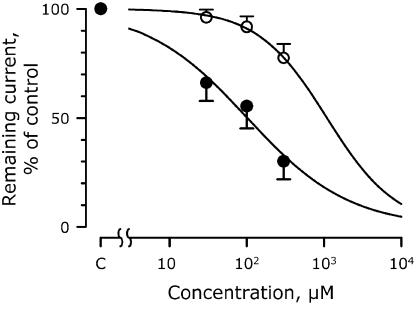
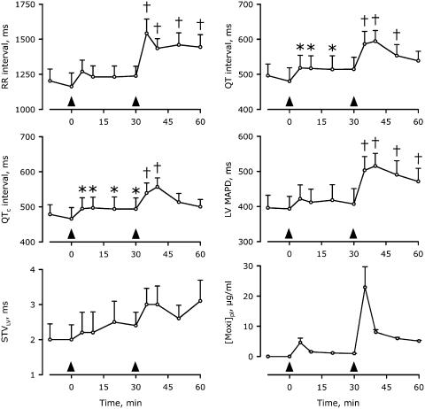
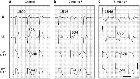
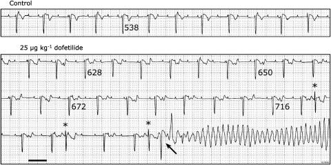
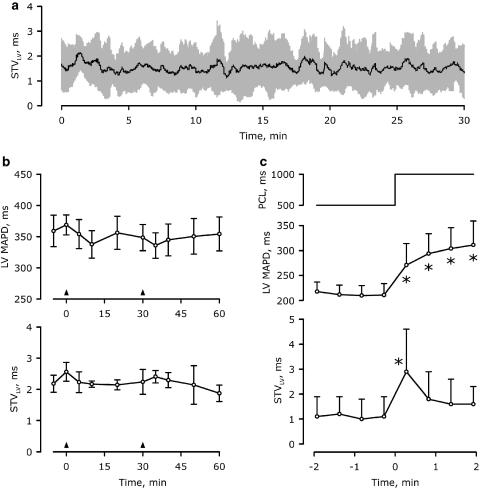
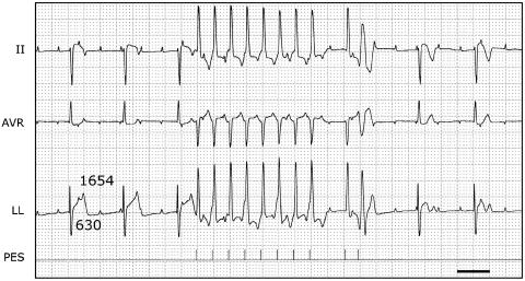
Experimental approach & key results: In transfected HEK cells, the IC(50)s for I (hERG) were 45+/-6 and 856+/-259 microg ml(-1) for moxifloxacin and azithromycin, respectively. Intravenous administration of 2 and 8 mg kg(-1) moxifloxacin (total peak-plasma concentrations 4.6+/-1.5 and 22.9+/-6.8 microg ml(-1)) prolonged the QT(c) in 6 anaesthetized dogs with chronic AV block by 7+/-3 and 21+/-19%, respectively. Similar intravenous doses of azithromycin (total peak-plasma concentrations 5.4+/-1.3 and 20.8+/-4.9 microg ml(-1)) had no electrophysiological effects in the same dogs. The reference compound, dofetilide (25 microg kg(-1) i.v.) caused QT(c) prolongation (29+/-15%) and TdP in all dogs. Beat-to-beat variability of repolarization (BVR), quantified as short-term variability of the left ventricular monophasic action potential duration, was only increased after dofetilide (1.8+/-0.7 to 3.8+/-1.5 ms; P<0.05).

Conclusion & implications: As neither moxifloxacin nor azithromycin caused TdP or an increase in the BVR, we conclude that both drugs can be used safely in clinical situations.


Figures

Figure 1
Figure 1
The blocking effects of moxifloxacin and azithromycin on IhERG expressed in HEK 293 cells. Data points represent the means of four cells. Concentration–response curves are shown, where the remaining current is plotted relative to its control level. Closed circles, moxifloxacin with an IC20 and IC50 of 12 and 102 μM, respectively, and a Hill coefficient of 0.66. Open circles, azithromycin with corresponding values of 265, 1091 and 0.97 μM.
Figure 2
Figure 2
Time-dependent electrophysiological effects of cumulative moxifloxacin administration in anaesthetized CAVB dogs. Arrowheads indicate the start of the 5-min infusion of 2 and 8 mg kg−1 moxifloxacin. [Moxi]pl, plasma concentration of moxifloxacin. *P<0.05 versus control (−10 or 0 min); P<0.05 versus control and 30 min. Mean±s.e. are shown. All plasma concentrations were significantly different from control (not shown). n=6 anaesthetized CAVB dogs.
Figure 3
Figure 3
Electrophysiological effects of cumulative doses of moxifloxacin in a representative anaesthetized dog with CAVB. Two ECG leads, LV and RV MAP recordings are shown in each panel. RR intervals are above lead II and QT times above lead LL. MAPDs are next to the action potentials. ECG calibrated to 1 mV cm−1. Scale bar, 20 mV on the MAP signals. Horizontal scale bar, 1 s. (a) Control situation before moxifloxacin administration. (b) 10 min after start of 2 mg kg−1 moxifloxacin infusion. (c) 10 min after start of 8 mg kg−1 moxifloxacin infusion. The low dose causes a change in the T-wave morphology, whereas the high dose alters the ventricular origin of activation. No TdP was observed with any dose of moxifloxacin. In this example, the plasma concentrations measured at the time of these tracings were 1.6 and 8.6 μg ml−1 after 2 and 8 mg kg−1, respectively.
Figure 4
Figure 4
Representative examples of the electrophysiological and proarrhythmic effects of dofetilide administration in an anaesthetized CAVB dog. Same dog as in Figure 3. Lead II is shown at control and during dofetilide administration (25 μg kg−1). TdP occurred 2.5 min after the start of administration. QT intervals are indicated below the respective T-wave. Three morphologically comparable single spontaneous extrasystoles (*) occur, the last one triggering a TdP (arrow). ECG calibrated to 1 mV cm−1. Horizontal scale bar, 1 s.
Figure 5
Figure 5
Representative examples of Poincaré plots obtained from the same dog used in the other experiments. Left panels are controls, whereas right panels are under the influence of various drugs. Thirty consecutive LV MAPDs are plotted against the value of the previous beat. Each plot is 200–500 by 200–500 ms. All control plots cluster close to the diagonal line. (a) 8 mg kg−1 moxifloxacin provokes prolongation of the LV MAPD but does not elevate BVR as the dimensions of the plot remain unchanged. (b) 8 mg kg−1 azithromycin has no effect on either LV MAPD or BVR. (c) 25 μg kg−1 dofetilide prolongs LV MAPD as well as increasing BVR as is clearly seen by the increased area of the Poincaré plot. Only dofetilide was associated with proarrhythmia.
Figure 6
Figure 6
Time-dependent behaviour of BVR. (a) Short-term variability calculation continuously for 30 min under control conditions (n=3). Black line, mean data; grey area, mean±s.d. (b) Combined data of the time-dependent electrophysiological effects of cumulative azithromycin administration in anaesthetized CAVB dogs (n=5). Arrowheads indicate the start of the 5-min infusion of 2 and 8 mg kg−1 azithromycin. No statistically significant changes were observed in either LV MAPD (upper panel) or STVLV (lower panel). Mean±s.e. are shown. (c) Dynamicity of LV MAPD and STVLV upon sudden changes in heart rate. Continuous pacing from the right ventricle at 500 ms cycle length for 2 min was followed by an instantaneous change in pacing cycle length (PCL) to 1000 ms (upper panel) for 2 min. LV MAPD and STVLV of the paced periods are depicted in the lower panels. *P<0.05 versus 500 ms PCL.
Figure 7
Figure 7
Pacing to induce proarrhythmia after administration of moxifloxacin. Three ECG leads (II, AVR and LL) are shown. After administration of 2 followed by 8 mg kg−1 moxifloxacin, the QT interval is 630 ms at a CL of the idioventricular rhythm of 1654 ms. A programmed electrical stimulation (PES) was performed, resulting in 8 beats with a CL of 500 ms, followed by a beat after 1200 ms and an extra beat after 400 ms. The extra beat was paced close to the refractory period as evidenced by the broad R wave. Neither this nor any other stimulation protocol lead to arrhythmias. Note, however, the different T-wave morphologies of the nonpaced beats before and after the stimulation protocol. Furthermore, a tendency towards T-wave alternans can be observed during pacing with a CL of 500 ms. Horizontal scale bar, 1 s.

References

    1. Anderson ME, Mazur A, Yang T, Roden DM. Potassium current antagonist properties and proarrhythmic consequences of quinolone antibiotics. J Pharmacol Exp Ther. 2001;296:806–810. - PubMed
    1. Andriole VT, Haverstock DC, Choudhri SH. Retrospective analysis of the safety profile of oral moxifloxacin in elderly patients enrolled in clinical trials. Drug Safety. 2005;28:443–452. - PubMed
    1. Antoons G, Stengl M, Thomsen MB, Beekman JD, Vos MA, Sipido KR. Sarcoplasmic reticulum Ca2+ release and repolarization lability in myocytes from the dog with chronic atrioventricular block (cAVB) Biophys J. 2005;88:298A.
    1. Antzelevitch C. Role of transmural dispersion of repolarization in the genesis of drug-induced torsades de pointes. Heart Rhythm. 2005;2:S9–S15. - PMC - PubMed
    1. Bischoff U, Schmidt C, Netzer R, Pongs O. Effects of fluoroquinolones on HERG currents. Eur J Pharmacol. 2000;406:341–343. - PubMed

MeSH terms