Review
. 2006 Dec;4(12):932-42.
doi: 10.1038/nrmicro1552. Epub 2006 Nov 6.

Group B Streptococcus: global incidence and vaccine development

Affiliations
Review

Group B Streptococcus: global incidence and vaccine development

Atul Kumar Johri et al. Nat Rev Microbiol. 2006 Dec.

Abstract

An ongoing public health challenge is to develop vaccines that are effective against infectious diseases that have global relevance. Vaccines against serotypes of group B Streptococcus (GBS) that are prevalent in the United States and Europe are not optimally efficacious against serotypes common to other parts of the world. New technologies and innovative approaches are being used to identify GBS antigens that overcome serotype-specificity and that could form the basis of a globally effective vaccine against this opportunistic pathogen. This Review highlights efforts towards this goal and describes a template that can be followed to develop vaccines against other bacterial pathogens.


Conflict of interest statement

Competing interests statement: The authors declare no competing financial interests.

Figures

Figure 1
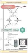
Figure 1. Approaches to vaccine development
A schematic demonstration of the essential steps required for vaccine development using the conventional approach (a), and reverse vaccinology (b). *Although DNA vaccines are potentially useful, in the case of Group B Streptococcus they have not been tested and are unlikely to be available in the short term. Reproduced with permission from REF. 15 © (2000) Elsevier.
Figure 2
Figure 2. The proteomic approach
A schematic representation of a proteomic approach to identify expressed proteins. Proteins are separated either by 2-dimensional electrophoresis or SDS-PAGE. The protein bands or spots are then excised, subjected to in-gel digestion with trypsin (a), and the resulting peptides are separated by high-performance liquid chromatography (HPLC) (b). The eluting peptides are ionized by electrospray ionization, enter the mass spectrometer (c), and are fragmented to collect sequence information (MS/MS (tandem mass spectroscopy) spectrum) (d). The amino-acid sequence of a peptide is obtained by comparing the MS/MS spectrum of the ionized peptide with predicted spectra generated from protein-sequence databases (e). Obtaining the peptide sequence allows identification of the original protein (f). Figure supplied courtesy of Steven Gygi, Harvard University, Boston.
Figure 3
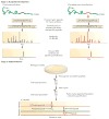
Figure 3. The ICAT strategy for quantifying differential protein expression
a | The ICAT reagent consists of three elements: an affinity tag (biotin) used to isolate ICAT-labelled peptides; a linker that can incorporate stable isotopes; and a reactive group with specificity toward thiol groups (cysteines). The reagent exists in two forms: heavy (contains eight deuterium atoms) and light (contains no deuterium atoms). b | Two protein mixtures of two different cell states (for example, a pathogen culture grown in vivo versus a culture grown in vitro) are treated with the isotopically light and heavy ICAT reagents, respectively; an ICAT reagent is covalently attached to each cysteinyl residue in every protein. Proteins from cell state 1 are shown in green, and proteins from cell state 2 are shown in blue. The protein mixtures are combined, digested to peptides, and ICAT-labelled peptides are isolated using the biotin tag. These peptides are separated by microcapillary high-performance liquid chromatography. The ratios of the original amounts of proteins from the two cell states are strictly maintained in the peptide fragments. The relative quantification is determined by the ratio of the peptide pairs. Every other scan is devoted to fragmenting and then recording sequence information about an eluting peptide by MS/MS (tandem mass spectroscopy). The protein is identified by searching the recorded sequence information against large protein databases. ICAT, isotope coded affinity tag chromatography; LC-MS/MS, liquid chromatography tandem mass spectroscopy. Reproduced with permission from Nature Biotechnology REF. 104 © (1999) Macmillan Publishers Ltd.
Figure 4
Figure 4. Quantification of proteins and phosphoproteins using the AQUA strategy
The AQUA strategy has two stages. Stage 1 involves the selection and standard synthesis of a peptide (or phosphopeptide (denoted here by pS)) from the protein of interest. During synthesis, stable isotopes are incorporated (for example, 13C, 15N) at a single amino-acid residue such as the leucine shown here (denoted by L*). These peptide internal standards (IS) are analysed by MS/MS (tandem mass spectroscopy) to examine peptide fragmentation patterns. The mass spectrometer is next set up to do a SRM (selected reaction monitoring) analysis in which a specific precursor-to-product ion transition is measured. Stage 2 is the implementation of the new peptide IS for precise sample protein quantification. Proteins are harvested from a biological sample (for example, bacteria isolated from the site of infection) and proteolysed with trypsin in the presence of the IS peptide and phosphopeptide. An LC–SRM (liquid chromatography–selected reaction monitoring) experiment then measures the abundance of a specific fragment ion from both the native peptide and the synthesized peptide as a function of reverse-phase chromatographic retention time. The absolute quantification of the protein of interest is determined by comparing the abundance of the known IS peptide with the native peptide. AQUA, absolute quantification. Reproduced with permission from REF. 105. © (2003) National Academy of Sciences, USA.

Similar articles

Cited by

References

    1. Schuchat A. Epidemiology of group B streptococcal disease in the United States: shifting paradigms. Clin Microbiol Rev. 1998;11:497–513. - PMC - PubMed
    1. Hansan SM, Uldbjerg N, Kilian M, Sorensen UB. Dynamics of Streptococcus agalactiae colonization in women during and after pregnancy and in their infants. J Clin Microbiol. 2004;42:83–89. - PMC - PubMed
    1. Dermer P, Lee C, Eggert J, Few B. A history of neonatal group B streptococcus with its related morbidity and mortality rates in the United States. J Pediatr Nurs. 2004;19:357–363. - PubMed
    1. Edwards MS, Baker CJ. Group B Streptococcal infections in elderly adults. Clin Infect Dis. 2005;41:839–847. - PubMed
    2. An excellent review providing in-depth information on GBS infection in elderly adults.

    1. Keefe GP. Streptococcus agalactiae mastitis: a review. Can Vet J. 1997;38:429–437. - PMC - PubMed

Publication types

MeSH terms