. 2006 Jul-Aug;12(7-8):127-36.
doi: 10.2119/2006-00024.Man.

Immunolocalization and expression of vascular endothelial growth factor receptors (VEGFRs) and neuropilins (NRPs) on keratinocytes in human epidermis

Affiliations
Free PMC article

Immunolocalization and expression of vascular endothelial growth factor receptors (VEGFRs) and neuropilins (NRPs) on keratinocytes in human epidermis

Xiao-Yong Man et al. Mol Med. 2006 Jul-Aug.
Free PMC article

Abstract

Vascular endothelial growth factor (VEGF) plays an important role in normal and pathological angiogenesis. VEGF receptors (VEGFRs, including VEGFR-1, VEGFR-2, and VEGFR-3) and neuropilins (NRPs, including NRP-1 and NRP-2) are high-affinity receptors for VEGF and are typically considered to be specific for endothelial cells. Here we showed expression of VEGFRs and NRPs on cultured epidermal keratinocytes at both mRNA and protein levels. We further localized these receptors by immunofluorescence (IF) staining in the epidermis of surgical skin specimens. We found positive staining for VEGFRs and NRPs in all layers of the epidermis except for the stratum corneum. VEGFR-1 and VEGFR-2 are primarily expressed on the cytoplasmic membrane of basal cells and the adjacent spinosum keratinocytes. All layers of the epidermis except for the horny cell layer demonstrated a uniform pattern of VEGFR-3, NRP-1, and NRP-2. Sections staining for NRP-1 and NRP-2 also showed diffuse intense fluorescence and were localized to the cell membrane and cytoplasm of keratinocytes. In another panel of experiments, keratinocytes were treated with different concentrations of VEGF, with or without VEGFR-2 neutralizing antibody in culture. VEGF enhanced the proliferation and migration of keratinocytes, and these effects were partially inhibited by pretreatment with VEGFR-2 neutralizing antibody. Adhesion of keratinocytes to type IV collagen-coated culture plates was decreased by VEGF treatment, but this reduction could be completely reversed by pretreatment with VEGFR-2 neutralizing antibody. Taken together, our results suggest that the expression of VEGFRs and NRPs on keratinocytes may constitute important regulators for its activity and may possibly be responsible for the autocrine signaling in the epidermis.


Figures

Figure 1
Figure 1
Expression of mRNA of VEGFRs and NRPs in cultured keratinocytes. RT-PCR analysis of VEGFR-1 (A), VEGFR-2 (B), VGFR-3 (E), NRP-1 (C), NRP-2 (F) and CD31 gene (H) in HUVECs (lane 1) and cultured keratinocytes from 3 different healthy samples (lanes 2–4). Lane 5 is a negative control showing no amplification from potential genomic DNA contamination. GAPDH served as an internal control for different amplification performed for respective genes (D, G, I).
Figure 2
Figure 2
Immunoblot analysis of VEGFRs and NRPs in cultured keratinocytes. The anti–VEGFR-1, anti–VEGFR-2, and anti–VEGFR-3 antibodies show bands at about 180 and 200 kDa. The anti–NRP-1 and anti–NRP-2 antibodies show bands at approximate 140 kDa. The anti-GAPDH antibody was used as loading control for normalization. M, marker; lane 1, HUVECs; lanes 2–4, cultured keratinocytes from 3 different healthy samples.
Figure 3
Figure 3
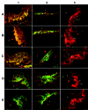
Representative immunofluorescent staining of VEGFRs and NRPs in human healthy skin epidermis. The presence of VEGFRs and NRPs is indicated by green fluorescence staining. Red indicates PI nuclear staining. Column 1, normal human skin from a 19-year-old male donor; column 2, human umbilical cord vein; column 3, negative staining by mouse IgG. The letters in the figures refer to epidermis (e), dermis (d), and basal layer of epidermis (b) in skin sections. The luminal side of the human umbilical cord vein is indicated by capital letter L. Scale bar, 50 μm. VEGFR-1 (A1) and VEGFR-2 (B1) are strongly detected at the stratum basal and spinosum adjacent to the basal of epidermis (arrows). VEGFR-3 (C1) is detected uniformly at all layers of the epidermis except for the horny cell layer (arrow). NRP-1 (D1) and NRP-2 (E1) are distributed uniformly among all layers of the epidermis except for the horny cell layer (arrows). Positive controls of human umbilical cord vein showed strong staining for HUVECs and smooth muscle cells for all 5 antibodies for VEGFRs and NRPs (arrows). Negative staining controls of human skin by using mouse IgG are unstained in all experiments (arrowheads refer to the outer edge of skin).
Figure 4
Figure 4
Effect of VEGF165 on keratinocyte proliferation. MTT-based assays were performed to determine the effects of VEGF alone or in combination with VEGFR-2 neutralizing antibody for 48-h treatment. Data represent the mean values of optical density measured in 6 wells for each treatment in a representative experiment. This experiment was repeated at least 3 times with similar results. **P ≤ 0.01; ***P < 0.001, significant differences between VEGF-treated and untreated samples. #P < 0.05, significant difference between VEGF-treated cells and cells treated with both VEGF and VEGFR-2 neutralizing antibody.
Figure 5
Figure 5
Effect of VEGF165 on keratinocyte migration. Incubation with VEGF165 significantly increases keratinocyte migration, and this induction is partially abrogated by pre-treatment with VEGFR-2 neutralizing antibody. Data are expressed as mean values ± SD for the total number of cells observed in 5 randomly selected fields from 3 separate wells. **P ≤ 0.01; ***P < 0.001, significant differences between VEGF-treated and untreated cells. ###P < 0.001, significant difference between VEGF-treated cells and cells treated with both VEGF and VEGFR-2 neutralizing antibody.
Figure 6
Figure 6
Effect of VEGF165 on keratinocyte adhesion. *P < 0.05; **P ≤ 0.01; ***P < 0.001, significant differences between VEGF-treated and untreated cells. Cells pretreated with VEGFR-2 neutralizing antibody had significantly increased keratinocyte adhesion compared with untreated cells (***P < 0.001). The reduction of adhesion of keratinocytes by VEGF treatment at 10 ng/mL was significantly restored by pretreatment with VEGFR-2 neutralizing antibody (###P < 0.001).

Similar articles

Cited by

References

    1. Ferrara N. Vascular endothelial growth factor: basic science and clinical progress. Endocr Rev. 2004;25:581–611. - PubMed
    1. Carmeliet P, Tessier-Lavigne M. Common mechanisms of nerve and blood vessel wiring. Nature. 2005;436:193–200. - PubMed
    1. Stacker SA, Achen MA. The vascular endothelial growth factor family: signalling for vascular development. Growth Factors. 1999;17:1–11. - PubMed
    1. Veikkola T, Alitalo K. VEGFs, receptors and angiogenesis. Semin Cancer Biol. 1999;9:211–20. - PubMed
    1. Neufeld G, Cohen T, Gengrinovitch S, Poltorak Z. Vascular endothelial growth factor (VEGF) and its receptors. FASEB J. 1999;13:9–22. - PubMed

Publication types

MeSH terms

Substances

LinkOut - more resources