. 2006 Dec;29(6):1349-57.

SPARC-induced migration of glioblastoma cell lines via uPA-uPAR signaling and activation of small GTPase RhoA

Affiliations

SPARC-induced migration of glioblastoma cell lines via uPA-uPAR signaling and activation of small GTPase RhoA

Sateesh Kunigal et al. Int J Oncol. 2006 Dec.

Abstract

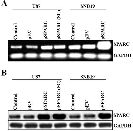
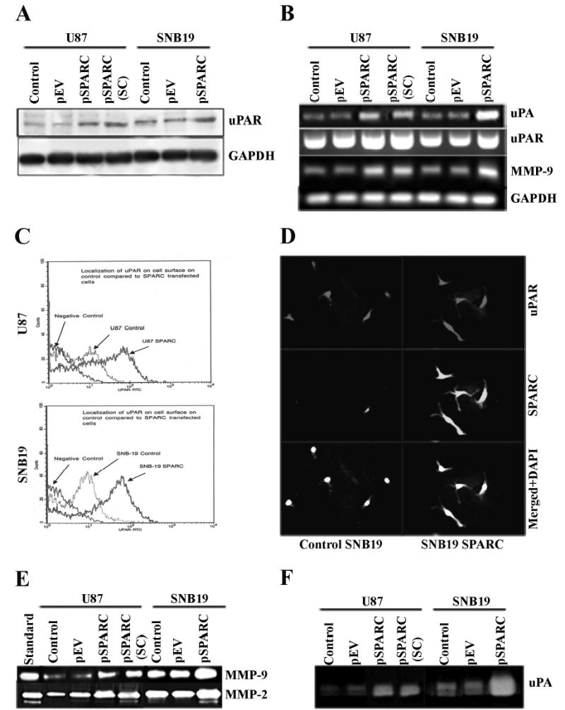
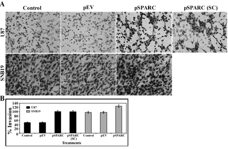
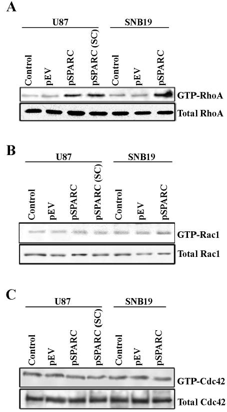
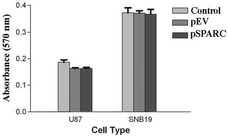
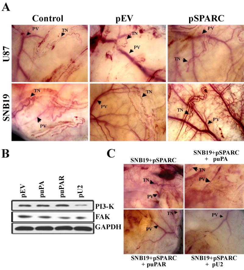
Secreted protein acidic and rich in cysteine (SPARC) is highly expressed in human gliomas where it promotes invasion and delays tumor growth, both in vitro and in vivo. SPARC, which interacts at the cell surface, has an impact on intracellular signaling and downstream gene expression changes, which might account for some of its effects on invasion and growth. Additionally in vitro studies demonstrated that SPARC delays growth, increases attachment, and modulates migration of tumor cells in an extracellular matrix-specific and concentration-dependent manner. Because the signaling aspect of this migration is neither well understood nor characterized, we overexpressed SPARC in both the minimally-invasive U87 cell line and in the most aggressive invasive cell line, SNB19. We first performed RT-PCR analysis and observed an upregulation of uPA and its receptor, uPAR. We also observed increased expression levels of matrix metalloproteinase-2 and -9 (MMP-2 and MMP-9). Western blot analysis confirmed these results, and the enzymatic activity of the metalloproteinases and uPA was further supported by zymography. Downstream of the uPA-uPAR interaction, upregulation of PI3-K occurred in cells overexpressing SPARC. Using GST-TRBD, we showed the upregulation of active GTP-bound RhoA, but neither Rac1 nor Cdc42 were activated. The inhibition of uPA and uPAR downregulated PI3-K activity and cell migration, as shown by matrigel invasion assay. A dorsal skin-fold chamber model revealed the high angiogenic activity of SPARC, though the proliferation of SPARC overexpressing cells was unaffected. Our results show that the small GTPase RhoA was a critical mediator of invasion or migration in the uPA-uPAR/PI3-K signaling pathway.


Figures

Figure 1
Figure 1. Overexpression of SPARC in the U87 and SNB19 Glioblastoma cells
U87 and SNB19 cells were transfected with pSPARC. After 48 h of incubation, (A) total RNA was extracted using TRIzol reagent, estimated and RT-PCR was performed for assessment of SPARC levels. Expression of GAPDH was verified for the uniform levels of cDNA. (B) SPARC levels were determined at the protein level by Western blot analysis using SPARC-specific antibodies. GAPDH was also immunodetected as a control to confirm equal loading of cell lysates. U87 SPARC (SC), a stable cell line, was included in both the cases in addition to the transfected sample.
Figure 2
Figure 2. Overexpression of SPARC increased expression of MMP-9, uPA and uPAR
U87 and SNB19 cells were transfected with pSPARC. After 48 h of incubation, (A) uPAR levels were determined at the protein level by Western blot analysis. GAPDH was also immunodetected as control to confirm equal loading of cell lysates. (B) Total RNA was extracted using TRIzol reagent, quantitated and RT-PCR was performed for assessment of uPA, uPAR and MMP-9 levels. Expression of GAPDH was verified for the uniform levels of cDNA. (C) After 48 h of incubation the cells were subjected to FACS analysis for uPAR expression. (D) Immunocytochemical analysis for the localization of uPAR and SPARC. (E & F) Activities of uPA and MMPs were analyzed by gelatin and fibrin zymography, respectively, by loading equal amounts of protein from the conditioned medium. U87 SPARC (SC), a stable cell line, was included in both the cases in addition to the transfected sample.
Figure 3
Figure 3. Overexpression of SPARC upregulates the invasive potential of SNB19 and U87 cells
U87 and SNB19 cells were transfected with pSPARC and incubated for 48 h. After the incubation period, the cells were trypsinized and washed once with serum-free medium. 1 × 10⁶ cells from each treatment were allowed to migrate through matrigel-coated transwell inserts (8-dm pores) for 24 h. The cells that invaded through the matrigel-coated inserts were stained (A) and photographed under a light microscope at 20× magnification and the percentage of invasion quantitated. (B) as described in the methods. Shown are the mean ± SD values from four separate experiments. U87 SPARC (SC), a stable cell line, was included in both cases in addition to the transfected sample.
Figure 4
Figure 4. Activation of RhoA but not Rac1 by SPARC
U87 and SNB19 cells were transfected with pSPARC. After a 48 h incubation period, cell lysates were incubated with the GST-TRBD and GST-PAK PBD beads and incubated for 45–60 min. The beads were then washed three times, resuspended in SDS loading buffer, and boiled for 10 min. SDS-PAGE and western blot analyses were performed. (A) Shows active RhoA; the lower panel shows the total RhoA in the control and treatments. (B) Shows active Rac1 and the total Rac1 in the control and treatments. (C) Shows active Cdc42 and total Cdc42 in the control and treatments. U87 SPARC (SC), a stable cell line, was included in both the cases in addition to the transfected sample.
Figure 5
Figure 5. SPARC has no effect on proliferation
1–2×10⁴ U87 and SNB19 cells were plated in microtiter plates, transfected with pSPARC, and incubated for 48 h. Then medium was removed from each well and 20 dL of 0.5 mg/ml MTT (3-4,5-dimethylthiazol-2-yl)-2,5-diphenyltetrazolium) in PBS was added to each well and allowed to incubate for a furth 4 h. 100 dL of DMSO was then added to each well to dissolve the formazan crystals. Absorbance values at 550 nm were measured with a microplate reader. These results were presented as percentage of cells treated with vehicle DMSO (0.25%). Shown are the ± SD values from four separate experiments (p<0.05).
Figure 6
Figure 6. Overexpession of SPARC enhances angiogenesis in gliomas
Enhancement of tumor angiogenesis in pSPARC vector was demonstrated by the mouse dorsal window assay. (A) U87 and SNB19 cells were transfected sith pSPARC and incubated for 48 h. After the incubation period, 1×10⁶ cells from each treatment condition were suspended in 100 dL of PBS, injected into diffuse chambers, and placed under the dorsal skin cavity in nude mice for 10 days. Then, the dorsal skin at the site of the diffuse chamber was removed, observed for neovascularization, and photographed. (PV = pre-existing vasculature; TN = tumor induced vasculature). (B) Involvement of the uPA-uPAR system in the signaling cascade SNB19 SPARC stable cell line was transfected with puPA, puPAR and pU2 and the levels of PI3-K, FAK and GAPDH determined by western blotting. (C) Inhibition of tumor angiogenesis in SNB19 SPARC stable cell line transfected with various constructs as shown by mouse dorsal window assay. (PV = pre-existing vasculature; TN = tumor induced vasculature).

Similar articles

Cited by

References

    1. Brekken RA, Sage EH. SPARC, a matricellular protein: at the crossroads of cell-matrix. Matrix Biol. 2000;19:569–580. - PubMed
    1. Golembieski WA, Rempel SA. cDNA array analysis of SPARC-modulated changes in glioma gene expression. J Neurooncol. 2002;60:213–226. - PubMed
    1. Yamanaka M, Kanda K, Li NC, Fukumori T, Oka N, Kanayama HO, Kagawa S. Analysis of the gene expression of SPARC and its prognostic value for bladder cancer. J Urol. 2001;166:2495–2499. - PubMed
    1. Sakai N, Baba M, Nagasima Y, Kato Y, Hirai K, Kondo K, Kobayashi K, Yoshida M, Kaneko S, Kishida T, Kawakami S, Hosaka M, Inayama Y, Yao M. SPARC expression in primary human renal cell carcinoma: upregulation of SPARC in sarcomatoid renal carcinoma. Hum Pathol. 2001;32:1064–1070. - PubMed
    1. Paley PJ, Goff BA, Gown AM, Greer BE, Sage EH. Alterations in SPARC and VEGF immunoreactivity in epithelial ovarian cancer. Gynecol Oncol. 2000;78:336–341. - PubMed

Publication types

MeSH terms