. 2006 Dec;29(6):1525-31.

Response of neuroblastoma cells to ionizing radiation: modulation of in vitro invasiveness and angiogenesis of human microvascular endothelial cells

Affiliations

Response of neuroblastoma cells to ionizing radiation: modulation of in vitro invasiveness and angiogenesis of human microvascular endothelial cells

Unmesh Jadhav et al. Int J Oncol. 2006 Dec.

Abstract

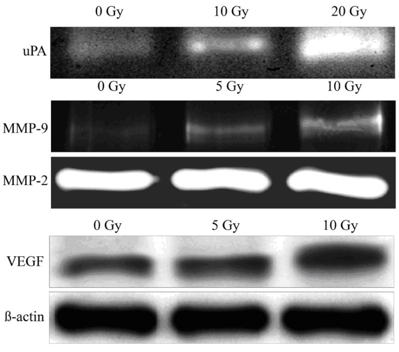
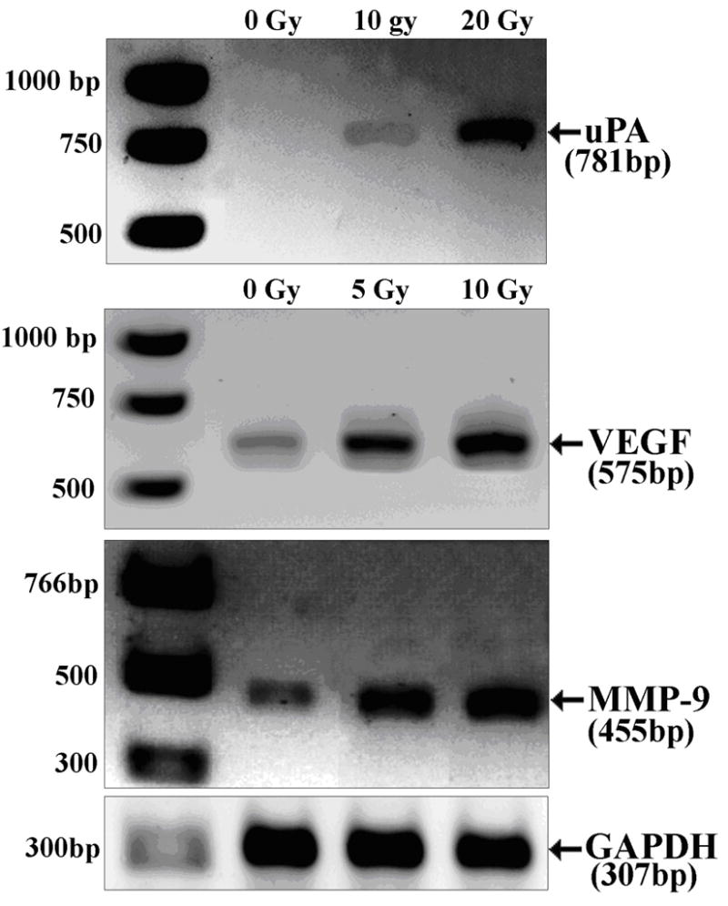
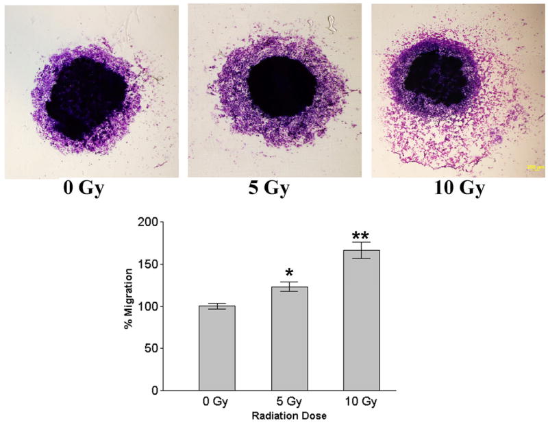
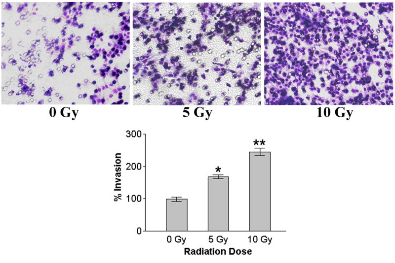
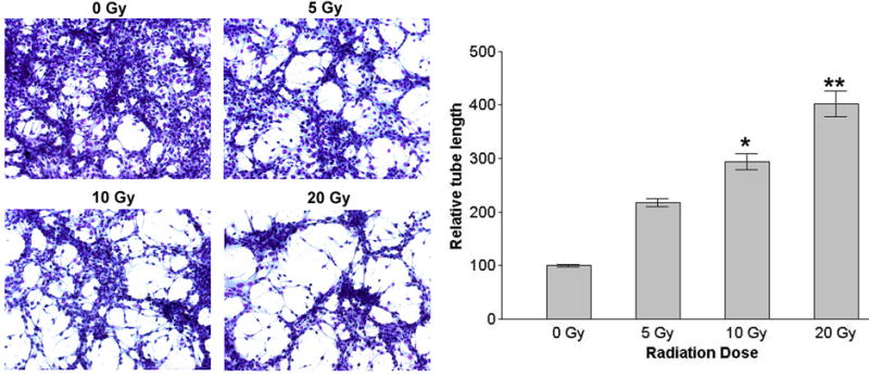
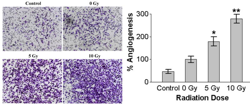
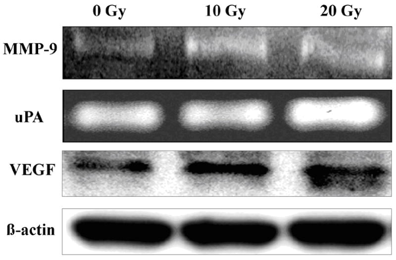
Neuroblastomas are the most common extra-cranial tumors of childhood and well known for their heterogeneous clinical behavior associated with certain genetic aberrations. Radiation therapy is an important modality for the treatment of high-risk neuroblastomas. In this study, we investigated whether ionizing irradiation modulate the migration and invasiveness of human neuroblastoma cells and expression of proangiogenic molecules known to be involved in tumor progression and metastasis. Irradiation of neuroblastoma cells resulted in increased migration and invasion as measured by spheroid migration and matrigel invasion assay respectively. Zymographic analysis revealed an increase in enzyme activity of MMP-9 and uPA in conditioned medium of irradiated neuroblastoma cells compared with non-irradiated cells. An increase in VEGF levels was also found in lysates of irradiated neuroblastoma cells. The up-regulation of uPA, MMP-9 and VEGF transcripts was also confirmed by RT-PCR analysis. Next, we examined the irradiated tumor cell-mediated modulation of endothelial cell behavior. Conditioned media from irradiated neuroblastoma cells enhanced capillary-like structure formation of microvascular endothelial cells. In a coculture system, irradiation of neuroblastoma cells enhanced endothelial cell invasiveness through Matrigel matrix. Endothelial cells treated with irradiated tumor cell conditioned medium were also analyzed for expression of uPA, MMP-9 and VEGF and compared to cells treated with non-irradiated tumor cell conditioned medium. These findings suggest that the irradiation effects of tumor cells could influence endothelial angiogenesis present in non-irradiated fields.


Figures

Figure 1
Figure 1
Effect of radiation on enzyme activity of uPA and MMP-9 and VEGF protein level in human neuroblastoma cells. Human neuroblastoma cells SK-N-AS were plated at equal numbers and irradiated with different radiation doses (10 Gy and 20 Gy). Serum-free conditioned medium was collected after 24 hr and electrophoresed under nonreducing conditions on 10% SDS-PAGE gels embedded with fibrinogen/plasminogen or gelatin. Gels were rinsed in 2.5% Triton X-100 for 30 min. at room temp and then incubated in 100mM glycine buffer pH 8.0 (for uPA zymography) or 50 mM Tris-HCl, 10mM CaCl2 buffer pH 7.6 (gelatin zymography) overnight at 37 0C. Gels were stained with Amido Black and areas of lysis were visualized as transparent bands. Irradiated and nonirradiated cells were extracted with lysis buffer containing protease inhibitors and electrophoresed on SDS-PAGE for analysis of VEGF and β-actin levels by immunoblotting.
Figure 2
Figure 2
Radiation induced changes in the transcripts of uPA, MMP-9 and VEGF in human neuroblastoma cells. Total RNA was extracted from irradiated and nonirradiated human neuroblastoma cells SK-N-AS and then reverse-transcribed using the cDNA cycle kit (Invitrogen). The cDNA was amplified with sense and antisense primers specific to uPA, MMP-9, VEGF and GAPDH. The amplified cDNA fragments were subjected to electrophoresis on a 1.5% agarose gel and photographed as ethidium bromide fluorescent bands. Densitometric values of uPA, MMP-9 and VEGF were normalized to GAPDH measurements.
Figure 3
Figure 3
Spheroid migration assay. Single multicellular spheroids of human neuroblastoma cells SK-N-AS were irradiated with different doses (5Gy and 10Gy) and cultured for 48 h. Then, spheroids were fixed and stained with Hema-3 and cellular migration from the spheroids was assessed using light microscopy. Values are mean ± SD of four determinations. * p<0.05; * *p<0.01, significantly different from non-irradiated cells.
Figure 4
Figure 4
Matrigel invasion assay. Irradiated and nonirradiated human neuroblastoma cells SK-N-AS were added to Matrigel-coated cell culture inserts in Transwell chambers. After 24 h incubation period, cells on the matrigel-coated side of the filter were removed with a cotton swab and the migrated cells remaining on the bottom part of the filters were fixed, stained, counted, photographed and the percentage of cells that had migrated through the Matrigel was determined. The graphical representation (bottom right panel) of invasion data as mean ± SD of two experiments done in duplicate. * p<0.05; * *p<0.01, significantly different from non-irradiated cells.
Figure 5
Figure 5
In vitro capillary-like structure formation. Capillary like structure formation in human microvascular endothelial cells (HMECs) was induced using conditioned medium from neuroblastoma tumor cells SK-N-AS. HMECs were allowed to grow in presence of conditioned media from irradiated and nonirradiated SK-N-AS cells for 16 h to measure the induction of cellular alignment into capillary-like structures. The graphical representative of three experiments done in duplicate expressed as mean ± SD. * p<0.05; * *p<0.01, significantly different from conditioned media of non-irradiated cells.
Figure 6
Figure 6
Endothelial Matrigel invasion assay. A modified coculture model of Matrigel invasion assay was performed to assess the effects on endothelial cells after selective radiation of tumor cells. Human neuroblastoma cells SK-N-AS were first seeded in 24-well plates. After irradiation of the neuroblastoma cells, Matrigel-coated 8-μm pore size transwell inserts with HMECs were added in the upper compartment and allowed to migrate toward the lower neuroblastoma compartment. After 24 of incubation, HMECs that had invaded into the underside of the membrane were fixed, stained and photographed. The graphical representation of invasion data as mean ± SD of two experiments done in duplicate. * p<0.05; * *p<0.01, significantly different from non-irradiated cells.
Figure 7
Figure 7
Effect of irradiated neuroblastoma tumor cell conditioned medium on the expression of proangiogenic molecules in human microvascular endothelial cells. Human microvascular endothelial cells (HMECs) were grown in conditioned medium from irradiated and nonirradiated human neuroblastoma cells SK-N-AS for 24 h. Serum free conditioned medium of HMECs was collected and analyzed for uPA and MMP-9 activity by substrate gel zymography. Cells were extracted in a lysis buffer containing protease inhibitors and analyzed by SDS-PAGE. VEGF protein levels were determined by immunoblotting with VEGF monoclonal antibodies (Santa Cruz Biotechnology). For detection, the ECL detection system (Amersham Biosciences) was used according to the manufacturer's instructions. Equal loading of the gels was confirmed by reincubation of the membrane with a monoclonal antibody for β-actin (Abcam).

Similar articles

Cited by

References

    1. Maris JM, Matthay KK. Molecular biology of neuroblastoma. J Clin Oncol. 1999;17:2264–2279. - PubMed
    1. Escobar MA, Grosfeld JL, Powell RL, West KW, Scherer LR, 3rd, Fallon RJ, Rescorla FJ. Long-term outcomes in patients with stage IV neuroblastoma. J Pediatr Surg. 2006;41:377–381. - PubMed
    1. Schmidt ML, Lukens JN, Seeger RC, Brodeur GM, Shimada H, Gerbing RB, Stram DO, Perez C, Haase GM, Matthay KK. Biologic factors determine prognosis in infants with stage IV neuroblastoma: A prospective Children's Cancer Group study. J Clin Oncol. 2000;18:1260–1268. - PubMed
    1. Laprie A, Michon J, Hartmann O, Munzer C, Leclair MD, Coze C, Valteau-Couanet D, Plantaz D, Carrie C, Habrand JL, Bergeron C, Chastagner P, Defachelles AS, Delattre O, Combaret V, Benard J, Perel Y, Gandemer V, Rubie H. Neuroblastoma Study Group of the French Society of Pediatric Oncology. High-dose chemotherapy followed by locoregional irradiation improves the outcome of patients with international neuroblastoma staging system Stage II and III neuroblastoma with MYCN amplification. Cancer. 2004;101:1081–1089. - PubMed
    1. Escobar MA, Grosfeld JL, Powell RL, West KW, Scherer LR, 3rd, Fallon RJ, Rescorla FJ. Long-term outcomes in patients with stage IV neuroblastoma. J Pediatr Surg. 2006;41:377–381. - PubMed

Publication types

MeSH terms