

Eye position-dependent activity in the primary visual area as revealed by fMRI
- PMID: 17089375
- PMCID: PMC6871435
- DOI: 10.1002/hbm.20296
Eye position-dependent activity in the primary visual area as revealed by fMRI
Abstract
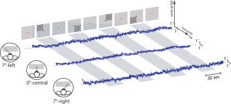
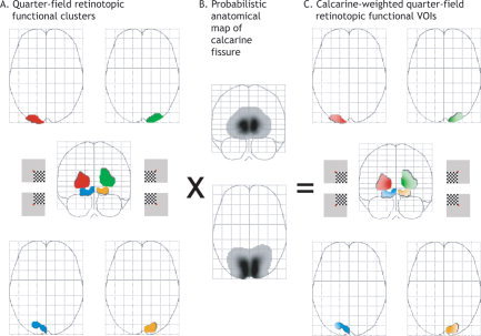
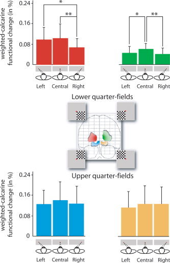
Internal senses of the position of the eye in the orbit may influence the cognitive processes that take into account gaze and limb positioning for movement or guiding actions. Neuroimaging studies have revealed eye position-dependent activity in the extrastriate visual, parietal, and frontal areas, but, at the earliest vision stage, the role of the primary visual area (V1) in these processes remains unclear. Functional MRI (fMRI) was used to investigate the effect of eye position on V1 activity evoked by a quarter-field stimulation using a visual checkerboard. We showed that the amplitude of V1 activity was modulated by the position of the eye, the activity being maximal when both the eye and head positions were aligned. Previous studies gave impetus to the emerging view that V1 activity is a cortical area in which contextual influences take place. The present study suggests that eye position may affect an early stage of visual processing.
Copyright 2007 Wiley-Liss, Inc.
Figures
References
-
- Andersen RA, Essick GK, Siegel RM 1985: Encoding of spatial location by posterior parietal neurons. Science 230: 456–458. - PubMed
-
- Andersen RA, Snyder LH, Li CS, Stricane B 1993: Coordinate transformations in the representation of spatial information. Curr Opin Neurobiol 3: 171–176. - PubMed
-
- Bilodeau L, Faubert J 1997: Isoluminance and chromatic motion perception throughout the visual field. Vision Res 37: 2073–2081. - PubMed
MeSH terms
Substances
LinkOut - more resources
Full Text Sources
Medical
