. 2006 Nov;4(11):e377.
doi: 10.1371/journal.pbio.0040377.

Assembly factor Omp85 recognizes its outer membrane protein substrates by a species-specific C-terminal motif

Affiliations

Assembly factor Omp85 recognizes its outer membrane protein substrates by a species-specific C-terminal motif

Viviane Robert et al. PLoS Biol. 2006 Nov.

Abstract

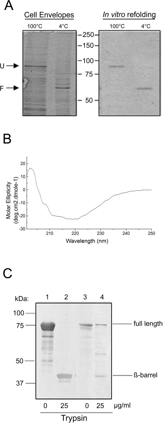
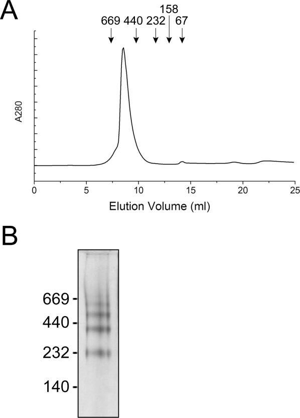
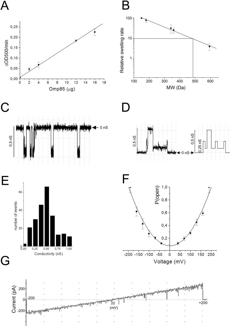
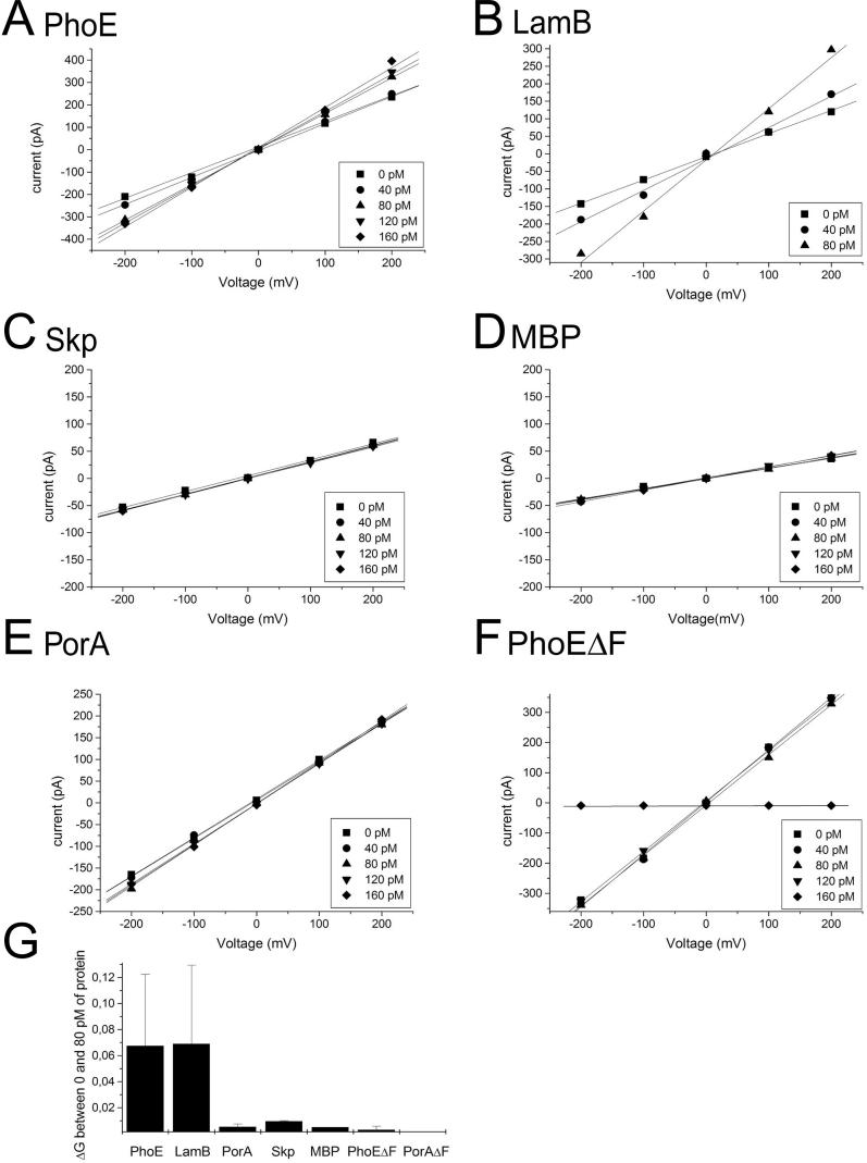
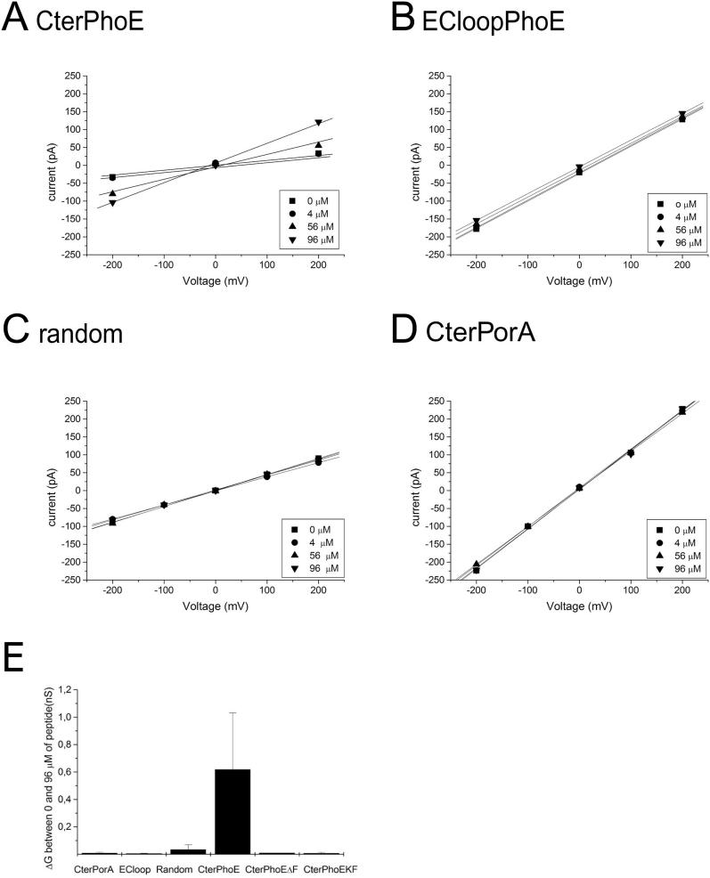
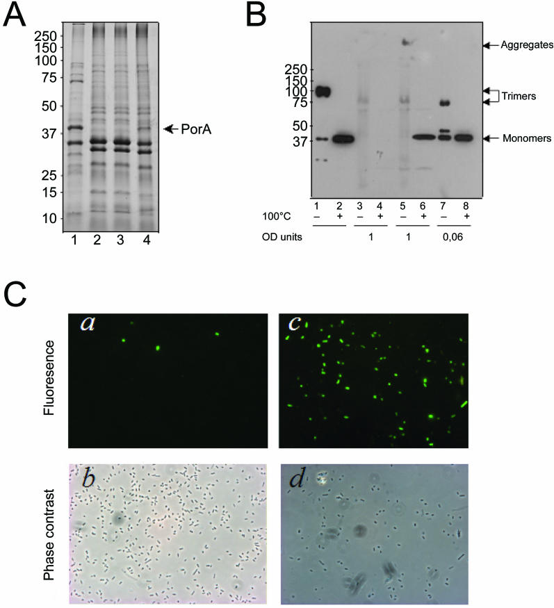
Integral beta-barrel proteins are found in the outer membranes of Gram-negative bacteria, mitochondria, and chloroplasts. The assembly of these proteins requires a proteinaceous apparatus of which Omp85 is an evolutionary conserved central component. To study its molecular mechanism, we have produced Omp85 from Escherichia coli in inclusion bodies and refolded it in vitro. The interaction of Omp85 with its substrate proteins was studied in lipid-bilayer experiments, where it formed channels. The properties of these channels were affected upon addition of unfolded outer-membrane proteins (OMPs) or synthetic peptides corresponding to their C-terminal signature sequences. The interaction exhibited species specificity, explaining the inefficient assembly of OMPs from Neisseria in E. coli. Accordingly, the in vivo assembly of the neisserial porin PorA into the E. coli outer membrane was accomplished after adapting its signature sequence. These results demonstrate that the Omp85 assembly machinery recognizes OMPs by virtue of their C-terminal signature sequence.


Conflict of interest statement

Competing interests. The authors have declared that no competing interests exist.

Figures

Figure 1
Figure 1. E. coli Omp85 Exhibits a β-Barrel Structure
(A) Comparison of the heat modifiability of in vitro folded Omp85 (right panel) and Omp85 from cell envelopes of E. coli strain AM1095 pCLyaeT (left panel) in SDS-PAGE. Samples were either heated to 100 °C or not before loading on the gel as indicated above the lanes. Only the relevant part of the gels is shown and the positions of molecular mass marker proteins are shown in the center (in KDa). F, folded Omp85; U, unfolded Omp85. (B) CD spectrum of Omp85. (C) Immunoblot showing tryptic fragments of refolded Omp85 (lanes 1 and 2) and TOP10F′ cell envelopes (lanes 3 and 4). The blot was probed with anti-Omp85 antiserum. The fragment indicated as β-barrel was N-terminally sequenced.
Figure 2
Figure 2. E. coli Omp85 Forms Multimers
(A) Size exclusion chromatography of purified refolded Omp85. Symbols indicate elution volumes of the MW standard proteins thyroglobulin (669 KDa), ferritine (440 KDa), catalase (232 KDa), aldolase (158 KDa), and ovalbumin (67 KDa) (Amersham Bioscience). (B) Blue Native PAGE gel of purified Omp85. The positions of molecular mass marker proteins are shown at the left (in KDa).
Figure 3
Figure 3. Pore-Forming Activity of Omp85
(A) Swelling rates of proteoliposomes reconstituted with the indicated amount of Omp85 in iso-osmotic solutions of L-arabinose. (B) Relative swelling rates of proteoliposomes containing Omp85 in solutions of sugars with different MW. The sugars used were arabinose (150 Da), glucose (180 Da), saccharose (342 Da), maltose (360 Da), and raffinose (594 Da). The data are shown relative to the swelling in arabinose and are the averages of at least four independent experiments. The indicated swelling rate corresponding to 10% of that in arabinose served to estimate the size of the channel. (C) Recording of Omp85 pores formed in planar lipid bilayers at an applied potential of −150 mV. (D) Current recordings showing sub-conductance states of Omp85 channels and its idealized current trace (right panel). The horizontal arrowhead shows the zero-conductance level. (E) Amplitude histogram of current derived from channel openings at +50, +100, and +150 mV. Results from four experiments were pooled. (F) Voltage dependence of the probability for the Omp85 channel of being in its open state. Data were normalized relative to the maximal mean current at 190 mV. Data points represent averages of two independent bilayers. (G) Voltage-ramp analysis of Omp85 channels from 0 to 200 mV and from 0 to −200 mV. In the experiment shown, eight 0.12-nS channels were present in the bilayer.
Figure 4
Figure 4. Denatured OMPs Affect Omp85 Pore Activity
(A–F) Voltage ramps were applied to bilayers containing several Omp85 channels before and after the addition of the proteins indicated above the panels. The amounts of protein added are shown in the inserts. The numbers of active 0.12-nS Omp85 channels in the experiments depicted were 10, 5, 3, 2, 7, and 13, in (A), (B), (C), (D), (E), and (F), respectively. (G) Increase in conductivity per Omp85 channel upon addition of 80 pM of each protein. Each experiment was repeated at least four times.
Figure 5
Figure 5. C-Terminal PhoE Peptide Affects Omp85 Pore Activity
(A–D) Voltage ramps were applied to bilayers containing Omp85 channels before and after the addition of the peptides shown above the panels. The amounts of peptides added are shown in the inserts. The numbers of active 0.12-nS Omp85 channels in the experiments depicted were 1, 6, 4, and 9, in (A), (B), (C), and (D), respectively. (E) Increase in conductivity per Omp85 channel upon addition of 96 μM of each peptide. Each experiment was repeated at least four times.
Figure 6
Figure 6. The Species Specificity of OMP Insertion between N. meningitidis and E. coli Is Related to the Nature of the C-Terminal Signature Sequence
(A) Coomassie-stained SDS-PAGE gel showing cell envelopes from N. meningitidis strain HB-1 (lane 1), and E. coli containing pCRII-TOPO (lane 2), pII-porAwt (lane 3), or pII-porA-Q (lane 4). (B) Cell envelopes were analyzed by SDS-PAGE after denaturation (+) or without denaturation (–) at 100 °C as indicated beneath the lanes. The gel was blotted and probed with anti-PorA antibody MN5C11G. Cell envelopes were from N. meningitidis strain HB-1 (lanes 1 and 2), and from E. coli carrying either pCRII-TOPO (lanes 3 and 4), pII-porAwt (lanes 5 and 6), or pII-porA-Q (lanes 7 and 8). Note the different loadings, which are indicated in optical density (OD) units of the original cultures below the blot. In (A) and (B), the E. coli strains were grown with 0.5 mM IPTG, and the positions of molecular mass marker proteins are shown at the left (in KDa). (C) Immunofluorescence microscopy analysis. The binding of a PorA-specific mAb to E. coli Top10F′ cells expressing wild-type PorA (a) or mutant PorA-Q (c) was evaluated. The corresponding phase-contrast images are shown in (b) and (d), respectively.

Comment in

References

    1. Koebnik R, Locher KP, Van Gelder P. Structure and function of bacterial outer membrane proteins: Barrels in a nutshell. Mol Microbiol. 2000;37:239–253. - PubMed
    1. Osborne AR, Rapoport TA, Van den Berg B. Protein translocation by the Sec61/SecY channel. Annu Rev Cell Dev Biol. 2005;21:529–550. - PubMed
    1. Eppens EF, Nouwen N, Tommassen J. Folding of a bacterial outer membrane protein during passage through the periplasm. EMBO J. 1997;16:4295–4301. - PMC - PubMed
    1. Lazar SW, Kolter R. SurA assists the folding of Escherichia coli outer membrane proteins. J Bacteriol. 1996;178:1770–1773. - PMC - PubMed
    1. Harms N, Koningstein G, Dontje W, Müller M, Oudega B, et al. The early interaction of the outer membrane protein PhoE with the periplasmic chaperone Skp occurs at the cytoplasmic membrane. J Biol Chem. 2001;276:18804–18811. - PubMed

Publication types

MeSH terms