Comparative Study
. 2006 Nov 7:6:52.
doi: 10.1186/1471-213X-6-52.

Comparative insights into questions of lepidopteran wing pattern homology

Affiliations
Comparative Study

Comparative insights into questions of lepidopteran wing pattern homology

Antónia Monteiro et al. BMC Dev Biol. .

Abstract

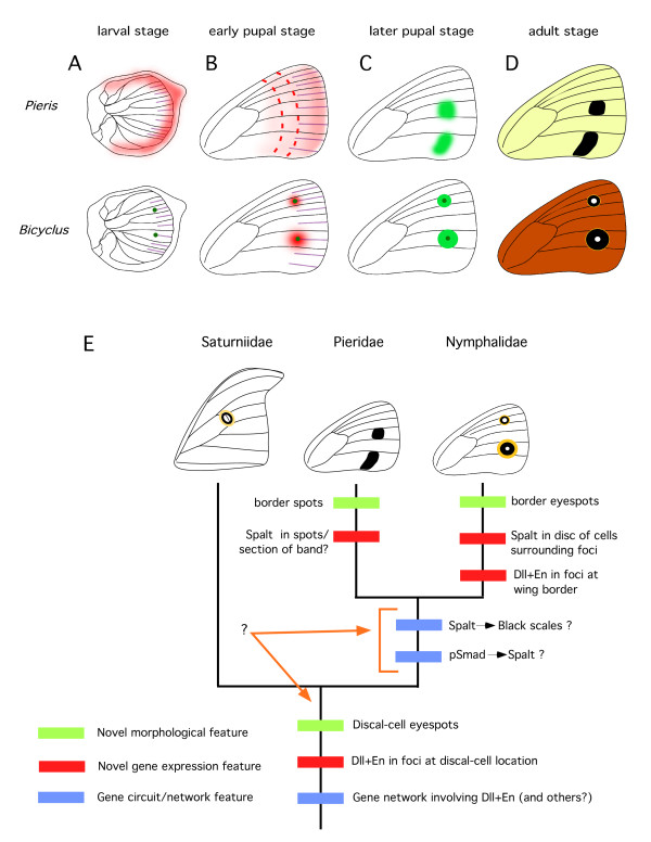
Background: Butterfly and moth eyespots can share a similar appearance, involving multiple concentric rings of colored scales, but usually occurring in non-homologous positions on the wing. Within the butterflies, on the other hand, spots that share the same homologous position may not share the concentric ring structure; and, in butterfly species that have eyespots with concentric rings, ectopic eyespots with a similar ring structure can be induced by means of a simple epidermal wound. The extent to which all these eyespots, natural or induced, share similar genes and developmental mechanisms is investigated here by means of protein in-situ localizations in selected butterfly and moth species. In addition to looking at some of the transcription factors previously identified as being involved in eyespot formation, we also tested the involvement of candidate genes from the Wingless and TGF-beta signaling pathways as putative morphogens for eyespot development.

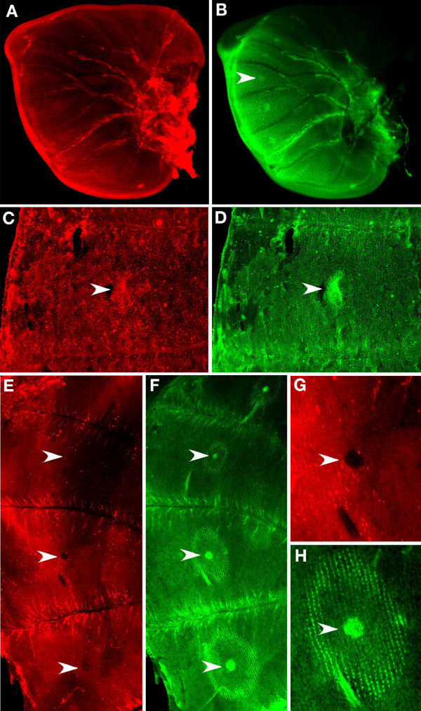
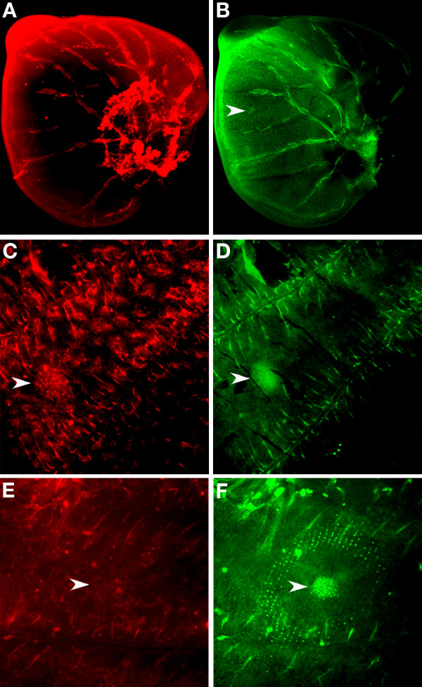
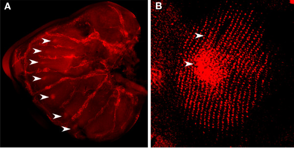
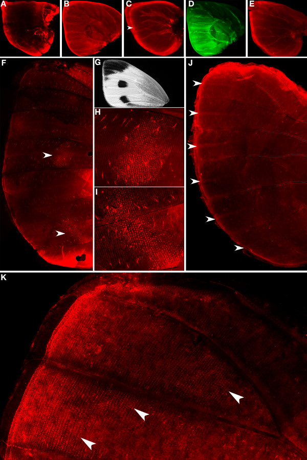
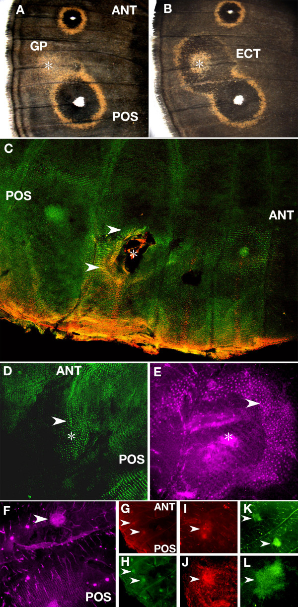
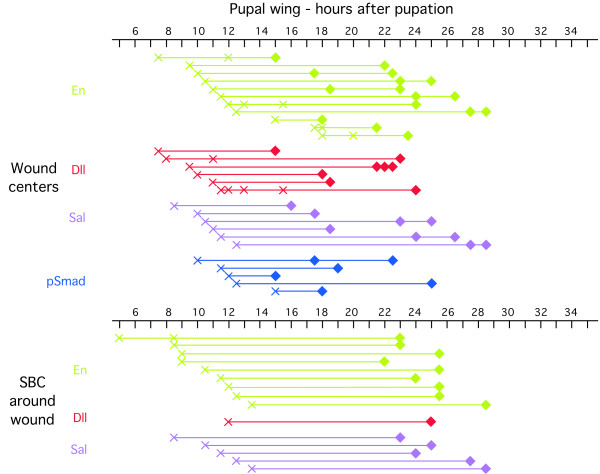
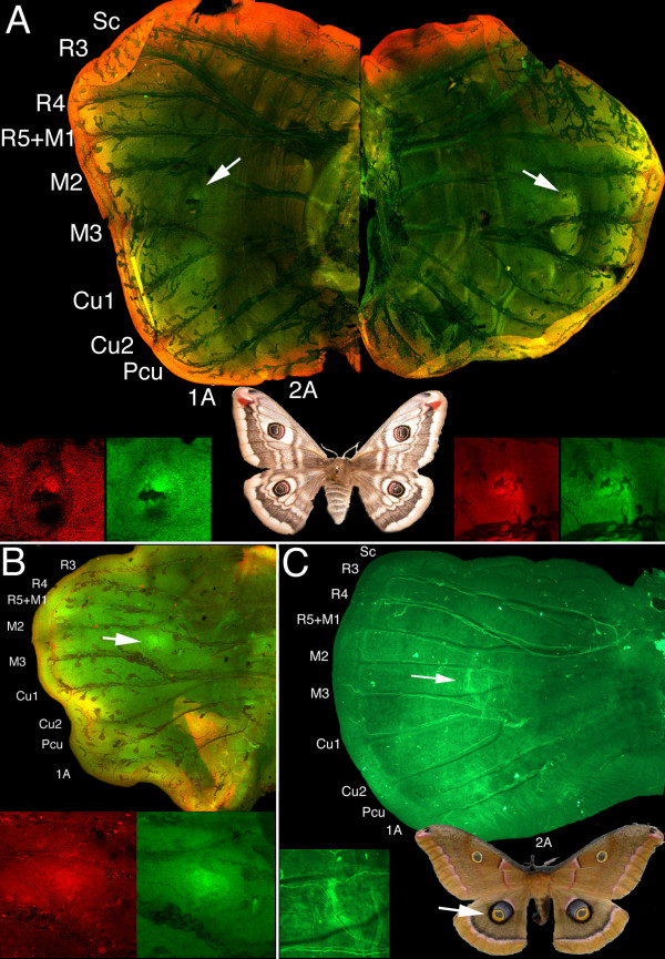
Results: Saturniid moth and nymphalid butterfly eyespots with concentric rings of color express at least two transcription factors, Distal-less and Engrailed, in the center of the future pattern. Nymphalid eyespots centers also express the ligand Wingless and an activated signal transducer, a phosphorylated Smad protein, but neither these proteins nor the previous two proteins are found in pierid spot centers, which consist of a single patch of color. Both butterfly wing patterns, however, express a third transcription factor, Spalt, a portion of whose expression domain maps to the black scales on the adult wing. Wounding a nymphalid wing, on the other hand, leads to upregulation of Distal-less, engrailed and spalt in subsets of cells around the wounding site, mimicking concentric eyespot development.

Conclusion: Wingless and TGF-beta ligands are both candidate morphogens involved in nymphalid butterfly eyespot formation. These eyespots, as well as saturniid moth eyespots with concentric circles, share two genes that are associated with the differentiation of the signaling cells in nymphalid eyespots. This commonality suggests that they may be produced via the same developmental mechanism despite their non-homologous location. By contrast, pierid butterfly spots of a single color share some of the same genes but appear to be produced by a different mechanism. Eyespots with concentric rings may have co-opted a wound healing genetic network during their evolution.


Figures

Figure 1
Figure 1
wingless expression in Bicyclus anynana larval and pupal wings. (A, B) Joint visualization of Wg (red) and En (green) in late 5th instar larval wing discs (50×); (A) Wg is present in the distal margin but not in the future eyespot centers in late 5th instar larval wing discs; (B) En is present in the future eyespot centers and in the posterior wing compartment; (C, D) Wg is present in the future eyespot centers at 12 h after pupation and its expression if coincident with that of En in the focus (large posterior forewing eyespot depicted) (100×); (E, F) Joint visualization of Wg and En at 16.5 h after pupation, while Wg levels (E) drop in the future eyespot centers, En (F) is still present in the future eyespot centers (50×); (G, H) Enlargement of 4th hindwing eyespot (200×). Arrowheads point to eyespot centers in all panels.
Figure 2
Figure 2
pSmad expression in Bicyclus anynana larval and pupal wings. (A, B) Joint visualization of pSmad (red) and En (green) in late 5th instar larval wing discs (50×); (A) pSmad is present along the margin but is absent in the future eyespot centers (arrowhead) where En (B) is visible; (C) pSmad and En (D) are present in the future eyespot centers, at 12 h after pupation. Note the patchiness of the pSamad staining outside of the focal area, perhaps indicating centers of epidermal cell growth (100×); (E) pSmad is no longer visible in the eyespot center (arrowhead) at 18 h after pupation, whereas en expression (F) extends to a ring pattern of scale-building cells surrounding the focus (200×).
Figure 3
Figure 3
Timing of visualizations of Dll, En, pSmad, Sal, and Wg in eyespot centers and scale-building cells (SBC) in larval and pupal wing discs. Each symbol along a row represents data for a unique individual, whereas symbols across rows may represent the same individual. Diamonds represent presence of the protein, zeroes represent absence of the protein, and circles with crosses represent absence of the protein below background levels. The second Wg row represents less common patterns to those observed and depicted in the first row, for the same time period.
Figure 4
Figure 4
spalt expression in Bicyclus anynana larval and pupal wings. (A) sal is expressed in the future eyespot centers (arrowheads), in the late 5th instar larval hindwing discs (50×); (B) sal expression extends to the scale-building cells in a larger circular pattern around the densely packed cells of the eyespot focus, at 19 h after pupation (arrowheads point to focus and to scale-building cells) (200×).
Figure 5
Figure 5
Expression of pSmad, wg, Dll, en, and sal in Pieris rapae larval and pupal wings. (A-E) Late 5th instar larval discs (50×); (A) pSmad is present in the distal margin; (B) Wg is also present in the distal margin and throughout the wing; (C) Dll is expressed in the distal margin and in intervenous stripes between wing veins (arrowhead); (D) en is expressed in the posterior compartment as well as along two intervenous stripes in the wing compartments that later carry the black spots; (E) sal is expressed in the distal margin; (F) sal is expressed in scale-building cells in the future black spots at 25.5 h after pupation (arrowheads) (100×); (G) Pieris rapae adult forewing showing two black spots and a black wing tip; (H) and (I) Enlargement of patches of sal expression from (F) (100×); (J) Dll is still expressed in intervenous stripes leading from the distal margin at 25.5 h after pupation (arrowheads) (100×). (K) Sal is present in scale-building cells at the distal tip of the wing at 26 h after pupation (arrowheads mark boundary of sal expression) (100×).
Figure 6
Figure 6
Gene expression after epidermal wounding in Bicyclus anynana. (A) Gold patches (GP) of scales are produced in some individuals around a wound site (star symbol) between the anterior (ANT) and posterior eyespots (POS), whereas (B) ectopic eyespots (ECT) containing both black scales and gold scales differentiate in other individuals; (C) Dll (red) and En (green) are present in scale-building cells around a wound site (yellow represents co-expression). Epidermis was wounded at 12 h after pupation (W = 12 h) followed by tissue fixation and protein visualization at 25 h after pupation (V = 25 h) (50×); (D) en is expressed in a patch of scale-building cells (arrowhead) around wound site (star symbol) (W = 9 h, V = 24 h) (100×); (E) sal is expressed in scale-building cells (arrowhead) around a wound site (approximate location shown by star symbol) (W = 13.5 h, V = 28.5 h) (200×). The absence of a continuous epidermis in the center of the wound site in (C) and (E) is the result of damage during the process of wing detachment from the overlying cuticle due to the presence of a wound scab; (F-L) Antibodies bind to the center of a wound (shown in all panels by arrowheads) in a non-specific fashion; (F) anti-Sal antibody binds to a cluster of non-differentiated cells (W = 11.5 h, V = 24 h) (100×); (G) Anti-Dll (W = 9 h, V = 24 h) (50×); (H) Anti-En (W = 9 h, V = 24 h) (50×). Note the two flanking eyespot foci on this wing (G, H) expressing Dll and en; (I) Anti-Wg (W = 9 h, V = 24 h) (100×); (J) Anti-pSmad (W = 9 h, V = 24 h) (100×); (K) Control staining using only an anti-mouse secondary antibody (and no primary antibody) also showing expression at the center of two wound sites (W = 9 h, V = 24 h) at 50X, and at 200X (L).
Figure 7
Figure 7
Timing of visualizations of antibodies targeting En, Dll, and Sal at wound centers and in scale-building cells surrounding wound centers after epidermal wounding. The age of the pupa when wounding was performed (cross symbol) is connected by an horizontal line to the age of the same pupa at the time the wing was extracted and fixed (diamond symbol), and stained with a particular antibody, Only wings where we observed some staining pattern are depicted in this figure. Data was condensed in the following way: more than one cross present on the same line represents observations in different individuals that were wounded at different times but whose wings were fixed at the same time after pupation. More than one diamond in the same line represent observations in different individuals that pupated at the same time and were fixed at different times. As explained in the text, the results for the wound centers (top part of the figure) may not represent real gene expression patterns, as non-specific antibodies were also shown to bind at these sites.
Figure 8
Figure 8
Expression of Dll and En in two saturniid species in the late larval wings, two to three days after the start of cocoon spinning. (A) Left and right forewings of Saturnia pavonia, showing Dll expressed in the margin (red) and also in future discal-eyespot centers (arrows and insets), together with En (green insets), two days after the beginning of cocoon spinning. The black marking next to the Dll- and En-expressing cells of the left wing appears to be a piece of debris, whereas the darker spots overlying these same cells on the right wing appear to be small pieces of trachea. (B) Hindwing of Saturnia pavonia showing an enlarged focus expressing Dll and En at the future discal-eyespot center. Note the absence of a cross vein at this stage. The area showing double staining in the bottom right sector of the wing represents a fold of the wing margin over part of the wing. (C) Hindwing of Antheraea polyphemus three days after beginning of cocoon spinning showing En present in a line marking the elongated central axis of the future discal-cell eyespot. In the pupal stages of both saturniids a cross vein will appear between M2 and M3, presumably at the position where En and Dll are expressed (see [42] for venation notation). All panels at 50× and insets at 100×.
Figure 9
Figure 9
Model for nymphalid eyespot and pierid spot development, and evolutionary hypothesis of spot/eyespot evolution. (A) During the late larval stage, Dll and Notch are expressed in several intervenous stripes (purple) midway between the wing veins in both B. anynana and P. rapae butterflies, but only a sub-set of wing cells express these and several other genes in enlarged foci at the end of those stripes, including sal (dark green; forewing represented here where only two foci differentiate). PSmad (red) is also present along the wing margin; B) In the early stages of pupal development, pSmad (red) is expressed in the future eyespot centers of B. anynana, but not in the spot centers of P. rapae. A gradient of this protein, however, may be present along the proximal-distal axis of the wing, being established from the marginal expression of TGF-β ligands in the late larval wings. This gradient could display a similar range of concentrations along bands spanning the anterior-posterior axis of the wing (red dashed lines); C) Later in pupal development while in B. anynana sal (light green) responds to pSmad levels generated from focal signaling, in P. rapae, sal is responding to particular pSmad levels expressed along the anterior-posterior axis (see text for further explanation of spot pattern). (D) The expression of sal determines where black scales will develop on the adult wing in both species. (E). Phylogenetic tree depicting a member from each lepidopteran family studied. The appearance of novel (non-homologous) features are depicted on the tree. These features can represent a novel phenotype, a novel gene expression pattern, or a novel gene circuit (see text for description). Note that additional information was used to map the origin of the discal-cell eyespots at the base of the tree, rather than in the branch representing the saturniid lineage, which would have been more parsimonious (see text).

References

    1. Wagner GP. The biological homology concept. Ann Rev Ecol Syst. 1989;20:51–69. doi: 10.1146/annurev.es.20.110189.000411. - DOI
    1. Nijhout HF. The development and evolution of butterfly wing patterns. Washington, Smithsonian Institution Press; 1991. p. 297.
    1. Kristensen NP. Lepidoptera, moths and butterflies: Evolution, systematics and biogeography. In: Kristensen NP, editor. Handbook of Zoology. Volume 1. New York , Walter de Gruyter; 1999.
    1. Abouheif E. Developmental genetics and homology: a hierarchical approach. Trends Ecol Evol. 1997;12:405–408. doi: 10.1016/S0169-5347(97)01125-7. - DOI - PubMed
    1. Gilbert SF, Bolker JA. Homologies of process and modular elements of embryonic construction. J Exp Zool. 2001;291:1–12. doi: 10.1002/jez.1. - DOI - PubMed

Publication types

Associated data

LinkOut - more resources