. 2007 Mar 15;109(6):2399-405.
doi: 10.1182/blood-2006-01-030643. Epub 2006 Nov 7.

CD34 cells from patients with trisomy 8 myelodysplastic syndrome (MDS) express early apoptotic markers but avoid programmed cell death by up-regulation of antiapoptotic proteins

Affiliations

CD34 cells from patients with trisomy 8 myelodysplastic syndrome (MDS) express early apoptotic markers but avoid programmed cell death by up-regulation of antiapoptotic proteins

Elaine M Sloand et al. Blood. .

Abstract

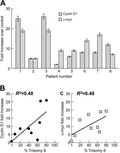
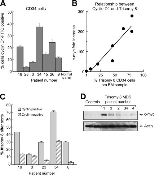
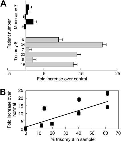
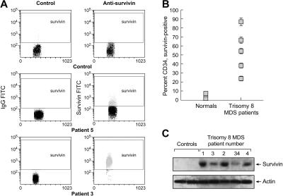
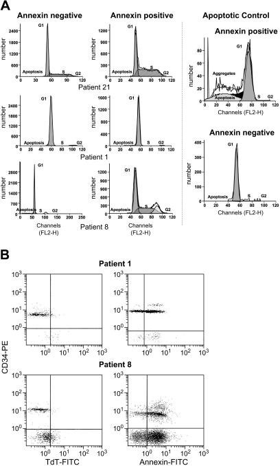
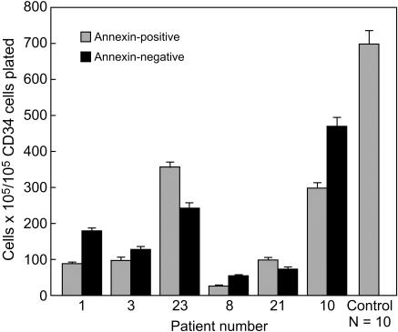
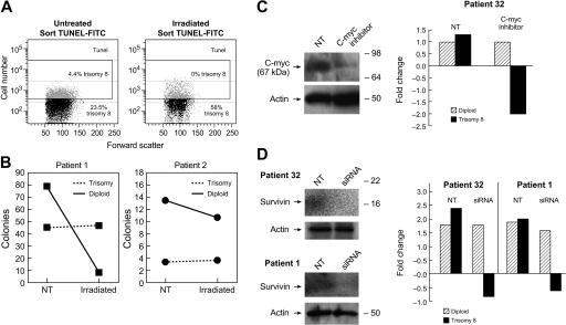
CD34 cells from patients with trisomy 8 myelodysplastic syndrome (MDS) are distinguished from other MDS cells and from normal hematopoietic cells by their pronounced expression of apoptotic markers. Paradoxically, trisomy 8 clones can persist in patients with bone marrow failure and expand following immunosuppression. We previously demonstrated up-regulation of c-myc and CD1 by microarray analysis. Here, we confirmed these findings by real-time polymerase chain reaction (PCR), demonstrated up-regulation of survivin, c-myc, and CD1 protein expression, and documented comparable colony formation by annexin(+) trisomy 8(-) CD34(+) and annexin(-) CD34 cells. There were low levels of DNA degradation in annexin(+) trisomy 8 CD34 cells, which were comparable with annexin(-) CD34 cells. Trisomy 8 cells were resistant to apoptosis induced by gamma irradiation. Knock-down of survivin by siRNA resulted in preferential loss of trisomy 8 cells. These results suggest that trisomy 8 cells undergo incomplete apoptosis and are nonetheless capable of colony formation and growth.


Figures

Figure 1
Figure 1
Up-regulation of CD1 and c-myc in CD34+ cells of patients with trisomy 8. CD34+ cells from the BM of patients with trisomy 8 were separated by flow cytometry, and mRNA was extracted and subjected to real-time PCR as described in “Materials and methods”; FISH was also performed to determine the percentage of trisomy 8 cells in the same sample. Both c-myc and CD1 m-RNAs were up-regulated in trisomy 8 compared with a control group of 10 healthy donors (A). The magnitude of increase in c-myc and CD1 expression was proportional to the number of trisomy 8 CD34 cells in the sample (B-C). Error bars represent standard error of the mean (SEM).
Figure 2
Figure 2
CD1 protein is overexpressed in trisomy 8. BMMNCs were stained with CD1-FITC and CD34-PE mAb and subjected to flow cytometry; CD34+ cells were gated, and the number of these cells staining with anti-CD1–FITC was quantitated. CD1 protein was increased in CD34 cells from patients with trisomy 8 compared with a group of healthy controls (A). CD1 expression was proportional to the number of trisomy 8 CD34 cells in the sample as determined by FISH using a specific chromosome 8 probe. (B) BMMNCs from patients with trisomy 8 were stained with CD34-PE and CD1-FITC. Cells were sorted into CD34+, CD1+, and CD1 aliquots by flow cytometry; FISH for chromosome 8 was performed on the sorted samples. There were increased trisomy 8 cells in the CD1+ fraction. (C) Immunoblots were performed on nuclear extracts of BMMNCs for c-myc. Increases in patients with trisomy 8 compared with healthy controls (n = 2) or other patients with MDS (n = 6) were seen. Error bars represent SEM.
Figure 3
Figure 3
Survivin, an IAP, is up-regulated in trisomy 8. RNA was extracted from purified CD34 cells with trisomy 8 and analyzed for survivin by real-time PCR; FISH was performed on sorted samples. Expression was compared with the mean of a panel of 10 healthy donors, as described in “Materials and methods.” Survivin mRNA was up-regulated in patients with trisomy 8 compared with controls and with a small group of equally cytopenic patients whose cytogenetics showed monosomy 7 (A). Expression was increased to a greater extent in samples with increased trisomy 8 cells (B). Error bars represent SEM.
Figure 4
Figure 4
Survivin protein is increased in trisomy 8 CD34 cells. Flow cytometry was performed on column-purified CD34 cells stained by intracellular staining as described in “Materials and methods” using survivin-FITC mAb. Survivin protein was increased in samples obtained from trisomy 8 BM (example seen in panel A). Entire sampling of patients examined is seen in panel B. Immunoblots were performed on total-cell lysates of BMMNCs for survivin. Increases in patients with trisomy 8 compared with healthy controls (n = 2) or other patients with MDS (n = 6) could be seen. Error bars represent SEM.
Figure 5
Figure 5
Absence of significant DNA degradation in annexin+ cells from patients with trisomy 8. (A) Cell-cycle analysis of sorted CD34 annexin+ and annexin cells showed that trisomy 8 cells were more heavily represented in the annexin+ fraction, but no significant numbers of cells with subgenomic DNA could be seen in this fraction (full dataset in Table 1). (B) When purified CD34 cells were stained with annexin-PE and CD34-FITC, or the TUNEL assay performed, there was significant annexin staining, but few cells were TUNEL+ (full dataset in Table 1).
Figure 6
Figure 6
Comparable hematopoietic colony formation by annexin+ and annexin CD34 cells from patients with trisomy 8. BMMNCs obtained from patients with significant trisomy 8 populations were stained with annexin-FITC and CD34-PE mAb and sorted into annexin+ and annexin aliquots of CD34 cells. These CD34 cells were plated in methycellulose for short-term culture as described in “Materials and methods.” Annexin+ CD34 cells were primarily trisomy 8 and gave rise to more hematopoietic colonies than did annexin cells in 3 of 4 patients. In all 5 samples obtained from healthy volunteers, the annexin+ CD34 cells failed to give rise to any colonies. FISH results on annexin+ and annexin fractions are listed in Table 1. Error bars represent SEM.
Figure 7
Figure 7
Resistance of trisomy 8 cells to apoptosis is abrogated by knock-down of c-myc and survivin.Trisomy 8 BM was subjected to 25 Gy of gamma radiation and cultured for 1 hour in media as described in “Materials and methods.” The TUNEL assay was performed to identify DNA nicks and TUNEL+ and TUNEL fractions sorted for both irradiated and nonirradiated fractions. The numbers of viable cells were decreased in the irradiated fraction, and the TUNEL+ cells increased from 18% to 30%. When FISH was performed on these fractions, trisomy 8 cells were consistently present in the TUNEL+ fraction (A). When control (nonirradiated) and irradiated lymphocyte-depleted BMMNCs were placed in short-term methylcellulose culture for 2 weeks the numbers of diploid colonies decreased to a greater extent than the numbers of trisomy 8 colonies (B). When an inhibitor of c-myc was cocultured with BMMNCs from a patient with trisomy 8, and immunoblotted, and FISH performed after 2 weeks of growth, there was a substantial decrease in the number of trisomy 8 cells coincident with a decrease in c-myc protein (C). BMMNCs transfected with siRNA specific for survivin were placed in hematopoietic growth factors for 36 hours at 37°C as described in “Materials and methods.” FISH and cell enumeration were performed before, and following culture using a labeled probe for chromosome 8. Immunoblotting was performed on total-cell lysates using antibody to survivin. (D) A substantial loss of trisomy 8 cells is seen compared with the baseline.

References

    1. Maciejewski J, Risitano A, Sloand EM, Nunez O, Young NS. Distinct clinical outcomes for cytogenetic abnormalities evolving from aplastic anemia. Blood. 2002;99:3129–3135. - PubMed
    1. Sloand EM, Mainwaring L, Fuhrer M, et al. Preferential suppression of trisomy 8 compared with normal hematopoietic cell growth by autologous lymphocytes in patients with trisomy 8 myelodysplastic syndrome. Blood. 2005;106:841–851. - PMC - PubMed
    1. Sloand EM, Kim S, Fuhrer M, et al. Fas-mediated apoptosis is important in regulating cell replication and death in trisomy 8 hematopoietic cells but not in cells with other cytogenetic abnormalities. Blood. 2002;100:4427–4432. - PubMed
    1. Maciejewski J, Selleri C, Anderson S, Young NS. Fas antigen expression on CD34+ human marrow cells is induced by interferon-gamma and tumor necrosis factor-alpha and potentiates cytokine-mediated hematopoietic suppression in vitro. Blood. 1995;85:3183–3190. - PubMed
    1. Michalopoulou S, Micheva I, Kouraklis-Symeonidis A, et al. Impaired clonogenic growth of myelodysplastic bone marrow progenitors in vitro is irrelevant to their apoptotic state. Leuk Res. 2004;28:805–812. - PubMed

MeSH terms