. 2006 Nov 14;103(46):17396-401.
doi: 10.1073/pnas.0608607103. Epub 2006 Nov 7.

Introduction of oncogenes into mammary glands in vivo with an avian retroviral vector initiates and promotes carcinogenesis in mouse models

Affiliations

Introduction of oncogenes into mammary glands in vivo with an avian retroviral vector initiates and promotes carcinogenesis in mouse models

Zhijun Du et al. Proc Natl Acad Sci U S A. .

Abstract

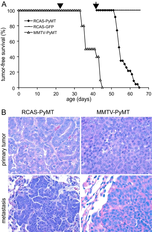
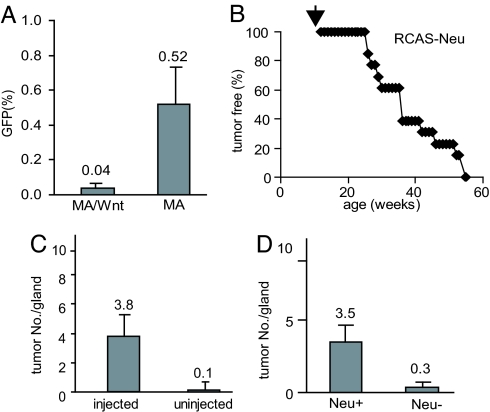
We have adapted the avian leukosis virus RCAS (replication-competent avian sarcoma-leukosis virus LTR splice acceptor)-mediated somatic gene transfer technique to introduce oncogenes into mammary cells in mice transgenic for the avian subgroup A receptor gene, tva, under control of the mouse mammary tumor virus (MMTV) promoter. Intraductal instillation of an RCAS vector carrying the polyoma middle T antigen (PyMT) gene (RCAS-PyMT) induced multiple, oligoclonal tumors within 3 weeks in infected mammary glands of MMTV-tva transgenic mice. The rapid appearance of these tumors from a relatively small pool of infected cells (estimated to be approximately 2 x 10(3) cells per gland by infection with RCAS carrying a GFP gene; RCAS-GFP) was accompanied by a high fraction of cells positive for Ki67, Cyclin D1, and c-Myc, implying strong proliferation competence. Furthermore, the tumors displayed greater cellular heterogeneity than did tumors arising in MMTV-PyMT mice, suggesting that RCAS-PyMT transforms a relatively immature cell type. Infection of mice transgenic for both MMTV-Wnt-1 and MMTV-tva with RCAS virus carrying an activated Neu oncogene dramatically enhanced tumor formation over what is observed in uninfected bitransgenic animals. We conclude that infection of mammary glands with retrovirus vectors is an efficient means to screen candidate oncogenes for their capacity to initiate or promote mammary carcinogenesis in the mouse.


Conflict of interest statement

The authors declare no conflict of interest.

Figures

Fig. 1.
Fig. 1.
Generation and infection of MMTV-tva transgenic mice. (A) Diagram of the MMTV-tva transgenic construct. (B and C) TVA is produced in mammary glands in MA mice. Immunohistochemical staining using rabbit antibodies against TVA was used to detect TVA in mammary sections from pubertal MA mice (B) and nontransgenic littermate controls (C). (D and E) TVA is produced in mammary epithelial cells in MA mice. Mammary sections from pubertal MA mice were stained for TVA (green) and Keratin 8 (red) (D) or for TVA (red) and α-SMA (green) (E). (F and G) Mammary cells from pubertal MA mice are susceptible to infection by RCAS. Primary mammary cells from MA mice were infected with RCAS-GFP viruses, stained for Keratin 8 (red) 3 days later, and photomicrographed (F). Mock infection (G) was included as a negative control. (H and I) Mammary cells in MA mice can be infected by intraductal delivery of RCAS. RCAS-GFP viruses were injected via the nipple duct into pubertal MA mice (H) and littermate nontransgenic controls (I). Mammary glands were collected 4 days later and stained for GFP by immunohistochemistry. Original images were captured with a ×40 objective.
Fig. 2.
Fig. 2.
RCAS-PyMT induces rapid formation of mammary tumors in MA mice. (A) RCAS-PyMT or RCAS-GFP was injected via the nipple duct into left no. 2–4 mammary glands (107 units per gland) in 13 and 10 6-week-old MA females, respectively. The infected mice were monitored for tumor appearance by palpation. The time of infection is noted by an arrow on the Kaplan-Meier tumor-free survival curves. The survival curve of line 634 of MMTV-PyMT transgenic mice (n = 10) is included for comparison, and the approximate time of puberty onset is indicated by an arrowhead. (B) Representative photomicrographs are shown for H & E-stained mammary tumors and lung metastases induced by RCAS-PyMT and from line 634 of MMTV-PyMT transgenic mice. A ×20 objective was used.
Fig. 3.
Fig. 3.
Use of the RCAS-mediated gene transfer method for identifying collaborating oncogenes. (A) Influence of the MMTV-Wnt-1 transgene on infection efficiency. Five 12-week-old mice bitransgenic for MMTV-tva and MMTV-Wnt-1 (MA/Wnt) were injected with RCAS-GFP viruses (107 units per gland). Four days after the infection, the fraction of GFP+ cells was quantified by flow cytometry. For comparison, five MA mice were also infected with RCAS-GFP; these mice were injected s.c. with estrogen (1 μg per mouse) and progesterone (1 mg per mouse) for 5 consecutive days starting 1 day before the viral infection to simulate the hyperplasia observed in MA/Wnt mice. (B) Tumor-free survival curve in MA mice infected by RCAS-Neu. Fourteen 12-week-old MA mice were stimulated with hormones as above, and six glands in each mouse were infected by RCAS-Neu (106 units per gland). Their tumor-free survival was monitored by palpation. (C) Tumor induction in mice bitransgenic for MMTV-tva and MMTV-Wnt-1. Twenty-six glands in seven 12-week-old mice were injected with RCAS-Neu (106 units per gland). All infected mice were killed 8 weeks later, and their mammary glands were examined grossly and microscopically to quantify the number of gross and occult tumors per infected or noninfected gland. (D) Quantitation of Neu+ and Neu tumors in infected glands. One section of each infected gland collected above was stained for HA to quantify the number of tumors expressing Neu.

References

    1. Green JE, Hudson T. Nat Rev Cancer. 2005;5:184–198. - PubMed
    1. Li Y, Hively WP, Varmus HE. Oncogene. 2000;19:1002–1009. - PubMed
    1. Fisher GH, Orsulic S, Holland E, Hively WP, Li Y, Lewis BC, Williams BO, Varmus HE. Oncogene. 1999;18:5253–5260. - PubMed
    1. Orsulic S. Mamm Genome. 2002;13:543–547. - PubMed
    1. Federspiel MJ, Bates P, Young JA, Varmus HE, Hughes SH. Proc Natl Acad Sci USA. 1994;91:11241–11245. - PMC - PubMed

Publication types

MeSH terms