. 2007 Feb;71(2):549-57.
doi: 10.1124/mol.106.028258. Epub 2006 Nov 7.

An opioid agonist that does not induce mu-opioid receptor--arrestin interactions or receptor internalization

Affiliations

An opioid agonist that does not induce mu-opioid receptor--arrestin interactions or receptor internalization

C E Groer et al. Mol Pharmacol. 2007 Feb.

Abstract

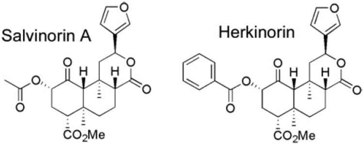
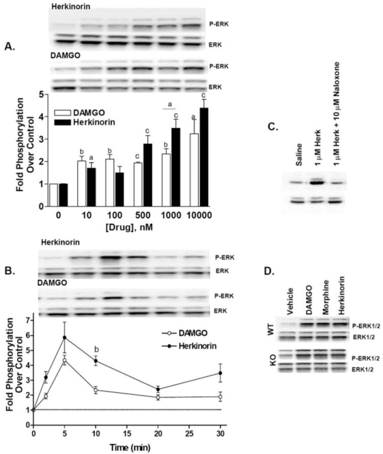
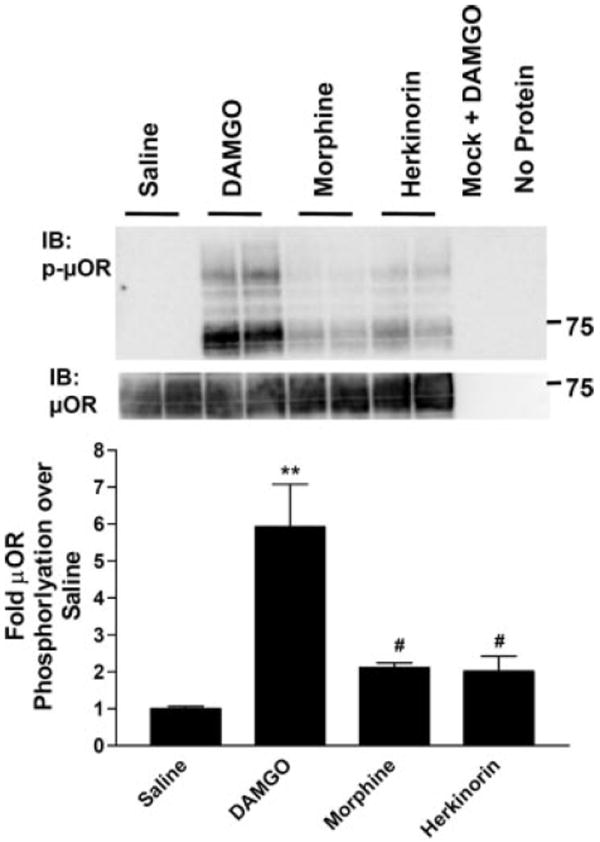
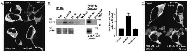
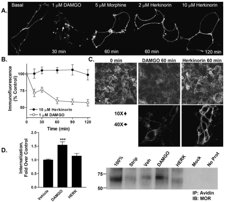
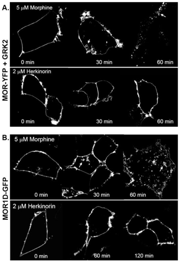
G protein-coupled receptor desensitization and trafficking are important regulators of opioid receptor signaling that can dictate overall drug responsiveness in vivo. Furthermore, different mu-opioid receptor (muOR) ligands can lead to varying degrees of receptor regulation, presumably because of distinct structural conformations conferred by agonist binding. For example, morphine binding produces a muOR with low affinity for beta-arrestin proteins and limited receptor internalization, whereas enkephalin analogs promote robust trafficking of both beta-arrestins and the receptors. Here, we evaluate muOR trafficking in response to activation by a novel mu-selective agonist derived from the naturally occurring plant product, salvinorin A. It is interesting that this compound, termed herkinorin, does not promote the recruitment of beta-arrestin-2 to the muOR and does not lead to receptor internalization. Moreover, whereas G protein-coupled receptor kinase overexpression can promote morphine-induced beta-arrestin interactions and muOR internalization, such manipulations do not promote herkinorin-induced trafficking. Studies in mice have shown that beta-arrestin-2 plays an important role in the development of morphine-induced tolerance, constipation, and respiratory depression. Therefore, drugs that can activate the receptor without recruiting the arrestins may be a promising step in the development of opiate analgesics that distinguish between agonist activity and receptor regulation and may ultimately lead to therapeutics designed to provide pain relief without the adverse side effects normally associated with the opiate narcotics.


Figures

Fig. 1
Fig. 1
Chemical structures of salvinorin A and herkinorin.
Fig. 2
Fig. 2
Herkinorin promotes ERK1/2 phosphorylation. Herkinorin and DAMGO induce ERK1/2 phosphorylation in HEK-293 cells stably expressing a hemagglutinin (HA-N terminus)-tagged mouse μOR. A, dose-response of herkinorin and DAMGO activation of ERK. Stimulation was for 10 min at the concentrations indicated. The dose-response comparison reveals a difference between DAMGO and herkinorin as assessed by two-way ANOVA: p < 0.05 (n = 2–6, n = 2 for 100 nM). Statistical legend: a, p < 0.05; b, p < 0.01; c, p < 0.0001 versus vehicle control, or between groups indicated by a bar, Student’s t test. B, time course of herkinorin- and DAMGO-induced phosphorylation of ERK. Cells were treated with 10 μM herkinorin or 1 μM DAMGO at the times indicated. Phopsho-ERK1/2 bands were analyzed by densitometry and normalized to total ERK levels (bottom bands) and densitometry is presented as fold stimulation (mean ± S.E.M.) over saline-treated controls. The time course comparison reveals a difference between DAMGO and herkinorin as assessed by two-way ANOVA, p < 0.05 (n = 3–5) and a significant difference between the compounds at the 10-min point. b, p < 0.01 Student’s t test, (n = 3–5). C, pretreatment with naloxone (10 μM) during the 30-min serum starvation blocked 1 μM herkinorin-induced ERK activation. D, activation of μOR in the absence of β-arrestins (βarr1&2-KO MEFs) still leads to ERK1/2 phosphorylation. Immunoblots were performed as described above using lysates from MEFs.
Fig. 3
Fig. 3
μ-Opioid receptor phosphorylation at serine 375 after agonist treatment. HEK-293 cells stably expressing μOR were treated with saline, 1 μM DAMGO, 10 μM morphine, or 10 μM herkinorin for 10 min. The receptor was immunoprecipitated from cell lysates using an anti-HA antibody-agarose bead complex. Representative Western blots using antibodies that recognize the μOR phosphorylated at serine 375 (top) or the total μOR (C-terminal antibody) from the same blot (bottom) are shown. Densitometric analyses of three independent experiments performed in duplicate or triplicate were normalized to total receptor per lane and expressed as fold stimulation over saline control for each blot. Data are presented as the mean ± S.E.M. **, p < 0.001; #, p < 0.01 versus DAMGO one-way ANOVA analysis of variance with Bonferroni’s multiple comparison test (n = 4–6).
Fig. 4
Fig. 4
Agonist-induced β-arrestin interactions with μOR in HEK-293 cells. HEK-293 cells transiently transfected with μOR and βarr2-GFP were imaged in real time after agonist treatment at room temperature. The cytosolic distribution of βarr2-GFP is shown in the untreated cells in the top left. A, βarr2-GFP translocation to μOR in HEK-293 cells. DAMGO (1 μM) treatment leads to βarr2-GFP translocation within 5 min (white arrow, punctate accumulation at membrane) whereas morphine (10 μM, 10 min) does not. Herkinorin (2 μM, 10 min) does not induce βarr2-GFP translocation. B, coimmunoprecipitation of β-arrestins and μOR after drug treatment. Cells were treated with vehicle (0.1% DMSO), 1 μM DAMGO, or 10 μM herkinorin for 5 min. Cells were then cross-linked using a cell-permeable cross-linking reagent (DSP). Cell lysates were immunoprecipitated by anti-HA-conjugated agarose beads, and proteins were resolved by SDS-PAGE under denaturing conditions. Blots (left), immunoblotting was performed using the β-arrestin antibody (A1CT); blots were stripped and then reprobed with the μOR antibody (Neuromics). Representative blots are shown. Mock refers to HEK cells transfected with empty vector. “No protein” contained no cell lysate in the immunoprecipitation. Antibody controls were performed on lysates run on the same gel as those shown for the immunoprecipitation. Densitometry (right), densitometry was measured from a total of three to four samples of each treatment prepared on 2 separate days. Shown are the means ± S.E.M. **, p < 0.01 versus WT or herkinorin (Herk), Student’s t test. C, βarr2-GFP translocation to μOR in HEK-293 cells overexpressing GRK2. Herkinorin does not promote βarr2-GFP translocation at 2 μM after 10 min or at 100 μM after 30 min. The same cells were treated with morphine (10 μM, 10 min) and βarr2-GFP translocates demonstrating that these cells do overexpress GRK2 because morphine does not induce visible translocation otherwise.
Fig. 5
Fig. 5
Agonist-induced internalization of μOR-YFP in HEK-293 cells. A, HEK-293 cells were transiently transfected with μOR (2 μg of cDNA) tagged at the C terminus with YFP. Cells were treated with the agonists indicated. Internalization can be seen after DAMGO treatment as indicated by the appearance of donut-like intracellular vesicles (white arrow) and the disappearance of membrane receptor localization as seen in the basal panel. Herkinorin and morphine do not induce receptor internalization. B, cell surface expression of receptor as determined by remaining N-terminal HA-immunoreactivity in nonpermeabilized cells after drug treatment. DAMGO, but not herkinorin, leads to a loss of cell surface expression after 60 to 120 min of drug treatment. Two-way ANOVA analysis reveals that the curves differ (p < 0.001) and that the DAMGO-treated cells display fewer surface receptors at each time point as determined by Bonferroni post hoc analysis (p < 0.001 at each time point after 0). C, immunocytochemistry of HA-MOR expression on cell surface after 60 min of agonist treatment. Top, 10× objective, images are taken directly from the 96-well plates assayed in 5B. Bottom, 40× objective images in parallel dishes. D, internalization of receptors as determined by cell surface biotinylation assay. Right blot, cell surface proteins were biotinylated and treated with indicated drug or vehicle (vehicle, 0.1% DMSO; 1 μM DAMGO or 10 μM herkinorin) for 1 h. Remaining surface proteins were then subjected to glutathione cleavage of biotin. Cells were then lysed, and remaining biotinylated proteins were immunoprecipitated with avidin-conjugated agarose beads. Proteins were resolved via SDS-PAGE under reducing conditions. Immunoblotting was performed with the C-terminal specific μOR antibody (Neuromics); 100% represents cells that were not stripped with glutathione; Strip represents remaining surface-biotinylated μOR after glutathione treatment and without drug treatment. Mock represents DAMGO treated HEK-293 cells that were transfected with empty vector. No Prot represents no cell lysate present in the immunoprecipitation. Densitometry (left), densitometry was measured from a total of five to seven samples of each treatment prepared on 3 separate days. ***, p < 0.001 versus vehicle; DAMGO induced significantly more internalization than herkinorin, p < 0.05. Herkinorin did not differ from vehicle, p > 0.1; Student’s t test.
Fig. 6
Fig. 6
Receptor internalization under conditions in which morphine will internalize μOR. A, agonist-induced μOR-YFP internalization in cells overexpressing GRK2. Experiments were performed as described in Fig. 5A with the addition of cotransfecting 5 μg of GRK2 cDNA. Morphine induces a redistribution of membrane-associated μOR-YFP to intracellular vesicle (white arrows); herkinorin does not. B, agonist induced μOR1-d-GFP internalization. μOR1-d-GFP (2 μg of cDNA) was transiently transfected into HEK-293 cells. Morphine treatment internalizes the receptor as indicated by the appearance of intracellular vesicles (white arrows), whereas herkinorin does not. Experiments were performed on at least three separate transfections; representative cells are shown.

References

    1. Alvarez V, Arttamangkul S, Williams JT. A RAVE about opioid withdrawal. Neuron. 2001;32:761–763. - PubMed
    1. Bailey CP, Couch D, Johnson E, Griffiths K, Kelly E, Henderson G. Mu-opioid receptor desensitization in mature rat neurons: lack of interaction between DAMGO and morphine. J Neurosci. 2003;23:10515–10520. - PMC - PubMed
    1. Belcheva MM, Vogel Z, Ignatova E, Avidor-Reiss T, Zippel R, Levy R, Young EC, Barg J, Coscia CJ. Opioid modulation of extracellular signal-regulated protein kinase activity is ras-dependent and involves Gbetagamma subunits. J Neurochem. 1998;70:635–645. - PMC - PubMed
    1. Bohn LM, Belcheva MM, Coscia CJ. Mitogenic signaling via endogenous kappa-opioid receptors in C6 glioma cells: evidence for the involvement of protein kinase C and the mitogen-activated protein kinase signaling cascade. J Neurochem. 2000a;74:564–573. - PMC - PubMed
    1. Bohn LM, Dykstra LA, Lefkowitz RJ, Caron MG, Barak LS. Relative opioid efficacy is determined by the complements of the G protein-coupled receptor desensitization machinery. Mol Pharmacol. 2004a;66:106–112. - PubMed

Publication types