. 2008 Jan;29(1):129-47.
doi: 10.1016/j.neurobiolaging.2006.09.015. Epub 2006 Nov 7.

Aging does not alter the number or phenotype of putative stem/progenitor cells in the neurogenic region of the hippocampus

Affiliations

Aging does not alter the number or phenotype of putative stem/progenitor cells in the neurogenic region of the hippocampus

Bharathi Hattiangady et al. Neurobiol Aging. 2008 Jan.

Abstract

To investigate whether dramatically waned dentate neurogenesis during aging is linked to diminution in neural stem/progenitor cell (NSC) number, we counted cells immunopositive for Sox-2 (a putative marker of NSCs) in the subgranular zone (SGZ) of young, middle-aged and aged F344 rats. The young SGZ comprised approximately 50,000 Sox-2+ cells and this amount did not diminish with aging. Quantity of GFAP+ cells and vimentin+ radial glia also remained stable during aging in this region. Besides, in all age groups, analogous fractions of Sox-2+ cells expressed GFAP (astrocytes/NSCs), NG-2 (oligodendrocyte-progenitors/NSCs), vimentin (radial glia), S-100beta (astrocytes) and doublecortin (new neurons). Nevertheless, analyses of Sox-2+ cells with proliferative markers insinuated an increased quiescence of NSCs with aging. Moreover, the volume of rat-endothelial-cell-antigen-1+ capillaries (vascular-niches) within the SGZ exhibited an age-related decline, resulting in an increased expanse between NSCs and capillaries. Thus, decreased dentate neurogenesis during aging is not attributable to altered number or phenotype of NSCs. Instead, it appears to be an outcome of increased quiescence of NSCs due to changes in NSC milieu.


Conflict of interest statement

Disclosure statement: (a) We do not have any actual or potential conflicts of interest including any financial, personal or other relationships with other people or organizations within 3 years of beginning the work submitted that could inappropriately influence (bias) their work. (b) All animal experiments in this study were performed as per the animal protocol approved by the Institutional Animal Care and Use Committees of the Duke University Medical Center and the Durham Veterans Affairs Medical Center.

Figures

Fig. 1
Fig. 1
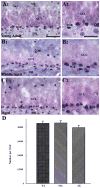
Distribution of Sox-2 immunoreactive cells in the dentate subgranular zone (SGZ) of young (A1), middle aged (B1) and aged (C1) male F344 rats. A2, B2 and C2 are magnified views of the SGZ from A1, B1 and C1. Note that the density of Sox-2 immunoreactive cells appears similar across the three age groups. The bar chart (D) compares the absolute numbers of Sox-2 immunoreactive cells in the SGZ of young (YA) middle-aged (MA) and aged (AG) rats. Note that the numbers of Sox-2 immunoreactive cells do not change with aging. DH, dentate hilus; GCL, granule cell layer; ML, molecular layer. Scale bar, A1, B1, and C1 = 50 μm; A2, B2, and C2 = 25 μm.
Fig. 2
Fig. 2
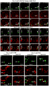
Sox-2 immunoreactive cells (in green color) in the subgranular zone (SGZ) of young and aged rats displaying different progenitor and glial cell markers (in red color), visualized by dual immunofluorescence and Z stacks using confocal microscopy. Examples include Sox-2 immunoreactive cells expressing GFAP (arrows in serial Z-sections, A1–J3), S-100β (arrows in K1–L3), NG2 (arrows in M1–N3). Arrowheads in all pictures denote Sox-2 immunoreactive cells lacking the expression of respective progenitor or glial cell markers. Scale bar, 25 μm.
Fig. 3
Fig. 3
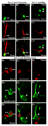
Parts (A1–B3) illustrate Sox-2 immunoreactive cells in the sub-granular zone (SGZ) of young and aged rats displaying vimentin (arrows), visualized by dual immunofluorescence and confocal microscopic analysis. Parts (C1–C3) illustrate a SGZ area containing Sox-2 immunoreactive cells (in green color) and rip positive oligodendrocytes (in red color). Note that Sox-2 immunoreactivity is absent within the nucleus of rip positive oligodendrocytes. Arrowheads denote Sox-2 immunoreactive cells lacking the expression of rip. Parts (D1–F3) illustrate examples of SGZ areas containing Sox-2 immunoreactive cells (in red color) and doublecortin immunopositive newly born neurons (in green color), visualized by dual immunofluorescence and confocal microscopy. Sox-2 immunoreactivity is present in only the doublecortin positive new neurons exhibiting no or primitive dendrites (arrows in D1–D3). In contrast, doublecortin positive new neurons with longer vertical dendrites (relatively mature new neurons) clearly lack Sox-2 expression (E1–F3). Arrowheads denote examples of doublecortin immunoreactive cells lacking Sox-2 expression in the subgranular zone. Scale bar, 25 μm.
Fig. 4
Fig. 4
Percentages of Sox-2 immunoreactive cells in the subgranular zone (SGZ) of young, middle-aged and aged hippocampus expressing GFAP, vimentin, S-100 or NG2. Note that the fractions of Sox-2 immunoreactive cells expressing GFAP, vimentin, S-100 and NG2 remain stable during the course of aging.
Fig. 5
Fig. 5
Sox-2 immunoreactive cells in the subgranular zone (SGZ) of young and aged rats displaying Ki67 (A1–B3) or BrdU (C1–D3), visualized by dual immunofluorescence and Z-section analyses using confocal microscopy. Note that all proliferating cells (as identified by the expression of Ki67 or BrdU) are positive for Sox-2 (arrows). Arrowheads denote Sox-2 immunoreactive cells lacking Ki67 or BrdU. Scale bar, 25 μm. Parts (E and F) illustrate the percentages of Sox-2 immunoreactive cells expressing Ki67 (E) and BrdU (F). Note that the percentages of Sox-2 immunoreactive cells expressing Ki67 or BrdU decline considerably during the course of aging. **p < 0.01; ***p < 0.001.
Fig. 6
Fig. 6
Distribution of GFAP+ astrocytes and their processes in the subgranular zone (SGZ) and granule cell layer (GCL) of young (A), middle aged (B) and aged (C) F344 rats. Parts (D–F) demonstrate the morphology of GFAP+ astrocytes encountered in the SGZ of young, middle-aged and aged rats, respectively. Asterisks denote nuclei of these cells. Scale bar, A–C = 50 μm; D–F, 10 μm. The bar chart in (G) illustrates the absolute number of GFAP+ cells within the SGZ of young (YA), middle-aged (MA) and aged (AG) rats. Note that, the numbers of GFAP+ astrocytes in the SGZ do not change with aging.
Fig. 7
Fig. 7
Distribution and morphology of vimentin+ radial glial cells in the subgranular zone (SGZ) and granule cell layer (GCL) of young (A1) middle aged (B1) and aged (C1) F344 rats. A2, B2 and C2 are magnified views of vimentin+ radial glia from the SGZ/GCL of three age groups of animals. Morphologically, radial glial cells in all three age groups exhibit a thick primary process that traverses radially through the GCL, bifurcates in the outer third of the GCL and branches in the dentate molecular layer. In addition, these radial glia also exhibit a few thinner processes that traverse the SGZ. Scale bar, A1–C1 = 100 μm; A2, B2, C2 = 25 μm. The bar chart in (D) illustrates the absolute number of vimentin+ radial glia in the SGZ of young (YA), middle-aged (MA) and aged (AG) rats. Note that, the numbers of vimentin+ cells in the SGZ do not change with aging.
Fig. 8
Fig. 8
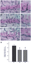
Distribution of RECA-1 immunoreactive capillaries in the dentate subgranular zone (SGZ) and granule cell layer of young (A1), middle-aged (B1) and aged (C1) F344 rats. A2, B2 and C2 illustrate magnified views of RECA-1 immunoreactive capillaries (presumably the vascular niches) in the SGZ of three age groups of animals. Note that the density of capillaries in the SGZ of the young hippocampus is greater than that in the middle-aged and aged hippocampus. Additionally, the capillaries in the aged hippocampus appear relatively wider than capillaries in the young hippocampus. Scale bar, A1, B1, and C1 = 100 μm; A2, B2, and C2 = 50 μm. The bar chart in (D) compares the volume fraction of RECA-1 immunoreactive capillaries/μm3 region of the SGZ between young (YA), middle-aged (MA) and aged (AG) groups. Note that, the overall volume of RECA-1 immunoreactive capillaries within the SGZ significantly decreases as early as middle age. GCL, granule cell layer. *p < 0.05.
Fig. 9
Fig. 9
Location of Sox-2 immunoreactive cells in relation to RECA-1 immunopositive capillaries in the subgranular zone (SGZ) of young (A1–A3), middle-aged (B1–B3) and aged (C1–C3) rats. Arrows denote Sox-2 immunoreactive cells that are located adjacent to RECA-1 immunopositive capillaries in the young, middle-aged, and aged SGZ. Arrowheads denote Sox-2 immunoreactive cells that are located away from the RECA-1 immunopositive capillaries. Scale bar, 25 μm. The bar chart in (D) compares the percentage of Sox-2 immunoreactive cells that are located adjacent to RECA-1 immunopositive capillaries in the SGZ of different age groups. Note that greater numbers of Sox-2 immunoreactive cells are located adjacent to RECA-1 immunopositive capillaries in the young SGZ, in comparison to both middle-aged and aged SGZ. *p < 0.05; ***p < 0.001.

References

    1. Altman J, Das GD. Autoradiographic and histological evidence of postnatal hippocampal neurogenesis in rats. J Comp Neurol. 1965;24:319–35. - PubMed
    1. Avilion AA, Nicolis SK, Pevny LH, Perez L, Vivian N, Lovell-Badge R. Multipotent cell lineages in early mouse development depend on SOX2 function. Genes Dev. 2003;17:126–40. - PMC - PubMed
    1. Bernal GM, Peterson DA. Neural stem cells as therapeutic agents for age-related brain repair. Aging Cell. 2004;3:345–51. - PubMed
    1. Bizon JL, Gallagher M. Production of new cells in the rat dentate gyrus over the lifespan: relation to cognitive decline. Eur J Neurosci. 2003;18:215–9. - PubMed
    1. Bizon JL, Lee HJ, Gallagher M. Neurogenesis in a rat model of age-related cognitive decline. Aging Cell. 2004;3:227–34. - PubMed

Publication types

MeSH terms