. 2007 Jan 5;282(1):345-52.
doi: 10.1074/jbc.M604503200. Epub 2006 Nov 8.

Disease-associated mutations at copper ligand histidine residues of superoxide dismutase 1 diminish the binding of copper and compromise dimer stability

Affiliations

Disease-associated mutations at copper ligand histidine residues of superoxide dismutase 1 diminish the binding of copper and compromise dimer stability

Jiou Wang et al. J Biol Chem. .

Abstract

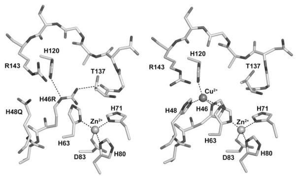
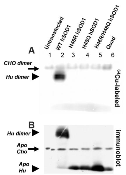
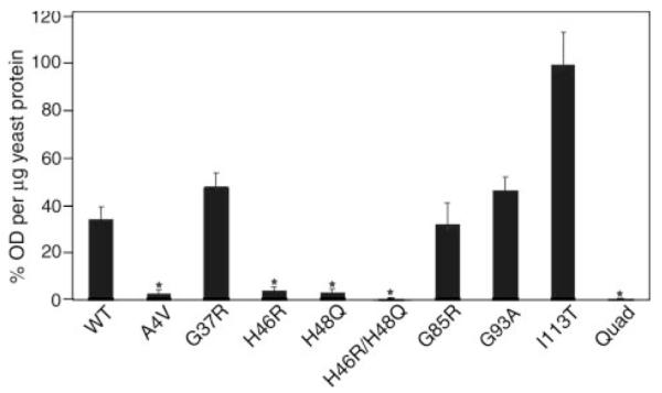
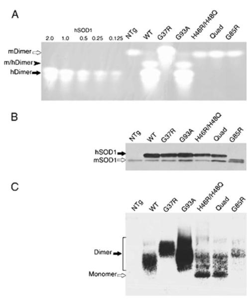
A subset of superoxide dismutase 1 (Cu/Zn-SOD1) mutants that cause familial amyotrophic lateral sclerosis (FALS) have heightened reactivity with (-)ONOO and H(2)O(2) in vitro. This reactivity requires a copper ion bound in the active site and is a suggested mechanism of motor neuron injury. However, we have found that transgenic mice that express SOD1-H46R/H48Q, which combines natural FALS mutations at ligands for copper and which is inactive, develop motor neuron disease. Using a direct radioactive copper incorporation assay in transfected cells and the established tools of single crystal x-ray diffraction, we now demonstrate that this variant does not stably bind copper. We find that single mutations at copper ligands, including H46R, H48Q, and a quadruple mutant H46R/H48Q/H63G/H120G, also diminish the binding of radioactive copper. Further, using native polyacrylamide gel electrophoresis and a yeast two-hybrid assay, the binding of copper was found to be related to the formation of the stable dimeric enzyme. Collectively, our data demonstrate a relationship between copper and assembly of SOD1 into stable dimers and also define disease-causing SOD1 mutants that are unlikely to robustly produce toxic radicals via copper-mediated chemistry.


Figures

FIGURE 1
FIGURE 1. The metal-binding sites of the human FALS SOD1 double mutant H46R/H48Q (left) and the wild-type enzyme (right)
The structure surrounding the copper-binding site of one subunit of SOD1-H46R/H48Q is compared with SOD1-wt (right). All subunits in the crystal of SOD1-H46R/H48Q showed perturbed copper-binding sites. The structure shown is that of the crystal subunits that most closely resemble SOD1-wt. Arg-143 and residues corresponding to metal ions ligands in the wild-type enzyme (metal ligand 46, 48, 63, 71, 80, 83, and 120) are labeled. The metal ions are represented by spheres. Metal ligand and hydrogen bonds are shown as dotted lines. In the left image, the side chain of the Arg residue substituted at position 46 donates a hydrogen bond to the carbonyl oxygen of Thr-137 on the opposite side of the active site channel, preventing the binding of copper ion (see text).
FIGURE 2
FIGURE 2. FALS mutations at copper-binding histidine residues of SOD1 dramatically reduce affinity for copper
A, CHO cells were transfected to express human SOD variants before being metabolically labeled with 50 μCi/ml of 64Cu for 3 h. 100 μg of each cell lysate was separated on a non-reducing 10% polyacrylamide gel containing 0.1% SDS. The 64Cu autoradiogram shows Cu-labeled endogenous hamster SOD1 dimer (solid arrow) in all samples, and Cu-labeled human SOD1 dimer (solid arrowhead) only in the WT sample. B, a duplicate of the gel used for 64Cu autoradiogram was analyzed by SOD1 immunoblot, which reveals endogenous hamster SOD1 monomer and human SOD1 monomers that are not labeled by 64Cu. Note: apo refers to the absence or presence of copper.
FIGURE 3
FIGURE 3. FALS mutations at copper-binding histidine residues of SOD1 dramatically reduce the strength of normal dimer interactions
Interactions between the human WT SOD1 and mutant subunits were measured by a yeast two-hybrid assay (see “Experimental Procedures”). Yeasts expressing mutant-SOD1 bait-fusion proteins were mated with yeasts expressing wild-type SOD1 target-fusion proteins. The interactions between two subunits were recorded by induction of a β-galactosidase reporter gene. Enzyme activity was measured by optical density values per microgram of yeast protein extract and then normalized totheI113T,whichhadthehighestlevels ofβ-galactosidase production. The datarepresentmeans±S.E.Threeor fourindependentbaitfusion yeast colonies, all of which express sufficient proteins to pass a repression assay, were used for each SOD1 variant (total number of measurements for each variant, n = 9–12). Significance levels were determined by one-way analysis of variance with Bonferroni corrections; the asterisks mark interaction measurements that are significantly lower than that of the wild-type to wild-type interaction values and mutant to wild-type interaction values with p < 0.01.
FIGURE 4
FIGURE 4. The electrophoretic migration of mutant SOD1 (H46R/H48Q and Quad-His) isolated from spinal cord resembles that of metal-deficient/reduced wild-type SOD1
Spinal cords from pre-symptomatic transgenic animals (2–3 months of age) were removed and homogenized in PBS by probe sonication for 30 s at 50% output (70 watts, Tekmar, Cincinnati, OH) and centrifuged at 100,000 × g for 5 min in a Beckman Airfuge (Beckman Coulter, Inc., Fullerton, CA). A, 100 μg of supernatant protein was separated by native gel electrophoresis and assayed for superoxide dismutase activity by gel assay as described under “Experimental Procedures.” Amounts (0.125–2.0 μg) of purified human SOD1 proteins were used as standards. Only dimeric holoenzymes show activity. In contrast to the WT, G37R, and G93A human SOD1 that show abundant active human holoenzymes (hDimer, solid arrow), H46R/H48Q, Quad, and G85R proteins show neither detectable activity nor affect the migration of mouse SOD1 homodimer (mDimer, open arrow), confirming their inability to form heterodimers (m/hDimer, solid arrowhead). NTg = non-transgenic sample. B, 0.5 μg of supernatant protein from mouse spinal cords was assayed for SOD1 protein levels by the standard SDS-PAGE using an antiserum against a conserved region in human SOD1 (hSOD1, solid arrow) and mouse SOD1 protein (mSOD1, open arrow). Note: G85R human protein runs slightly above the mouse SOD1. C, supernatant protein (5 μg) from mouse spinal cords was separated by native gel electrophoresis and immunoblotted using an antiserum that recognizes the human SOD1 but not the mouse protein. The homodimers (solid arrow) formed by WT, G37R, or G93A are consistent with those in A. The majorities of H46R/H48Q and Quad mutants migrate at positions similar to reduced and de-metallated hSOD1-WT (open arrow). The less abundant mutant G85R appears to migrate at a position expected for dimeric enzyme.

References

    1. Rosen DR, Siddique T, Patterson D, Figlewicz DA, Sapp P, Hentati A, Donaldson D, Goto J, O’Regan JP, Deng H-X, Rahmani Z, Krizus A, McKenna-Yasek D, Cayabyab A, Gaston SM, Berger R, Tanzi RE, Halperin JJ, Herzfeldt B, Van den Bergh R, Hung W-Y, Bird T, Deng G, Mulder DW, Smyth C, Laing NG, Soriano E, Pericak-Vance MA, Haines J, Rouleau GA, Gusella JS, Horvitz HR, Brown RH., Jr. Nature. 1993;362:59–62. - PubMed
    1. Andersen PM, Morita M, Brown RH., Jr. In: Amyotrophic Lateral Sclerosis. Brown RH Jr., Meininger V, Swash M, editors. Martin Dunitz Ltd.; London: 2000. pp. 223–250.
    1. Cleveland DW, Rothstein JD. Nat. Rev. Neurosci. 2001;2:806–819. - PubMed
    1. Hart PJ. Curr. Opin. Chem. Biol. 2006;10:131–138. - PubMed
    1. Borchelt DR, Lee MK, Slunt HH, Guarnieri M, Xu Z-S, Wong PC, Brown RH, Jr., Price DL, Sisodia SS, Cleveland DW. Proc. Natl. Acad. Sci. U. S. A. 1994;91:8292–8296. - PMC - PubMed

Publication types

Associated data

LinkOut - more resources