. 2006 Nov 21;103(47):17955-60.
doi: 10.1073/pnas.0606212103. Epub 2006 Nov 8.

Inhibition of dynamin completely blocks compensatory synaptic vesicle endocytosis

Affiliations

Inhibition of dynamin completely blocks compensatory synaptic vesicle endocytosis

A Jamila Newton et al. Proc Natl Acad Sci U S A. .

Abstract

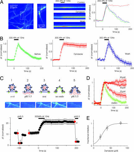
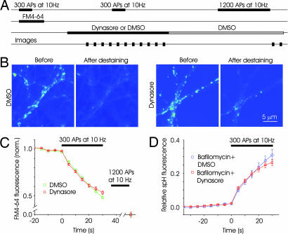
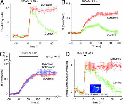
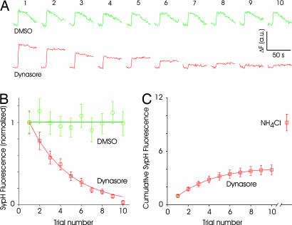
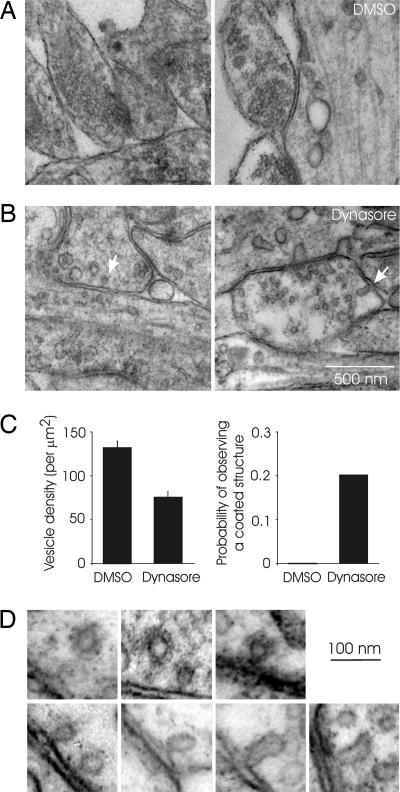
The ability of synapses to sustain signal propagation relies on rapid recycling of transmitter-containing presynaptic vesicles. Clathrin- and dynamin-mediated retrieval of vesicular membrane has an undisputed role in synaptic vesicle recycling. There is also evidence for other modes of vesicle retrieval, including bulk retrieval and the so-called kiss-and-run recycling. Whether dynamin in required for these other modes of synaptic vesicle endocytosis remains unclear. Here, we have tested the role of dynamin in synaptic vesicle endocytosis by using a small molecule called dynasore, which rapidly inhibits the GTPase activity of dynamin with high specificity. Endocytosis after sustained or brief stimuli was completely and reversibly blocked by dynasore in cultured hippocampal neurons expressing the fluorescent tracer synaptopHluorin. By contrast, dynasore had no effect on exocytosis. In the presence of dynasore, low-frequency stimulation led to sustained accumulation of synaptopHluorin and other vesicular proteins on the surface membrane at a rate predicted from net exocytosis. These vesicular components remained on surface membranes even after the stimulus was terminated, suggesting that all endocytic events rely on dynamin during low-frequency activity as well as in the period after it. Ultrastructural analysis revealed a reduction in the density of synaptic vesicles and the presence of endocytic structures only at synapses that were stimulated in the presence of dynasore. In sum, our data indicate that dynamin is essential for all forms of compensatory synaptic vesicle endocytosis including any kiss-and-run events.


Conflict of interest statement

The authors declare no conflict of interest.

Figures

Fig. 1.
Fig. 1.
Dynasore reversibly blocks endocytosis in hippocampal synapses. (A Left) Image of axons and presynaptic terminals labeled with spH. (A Center) Higher magnification image of a region from which a single bouton was chosen for linescan display. The line scan shows the time course of fluorescence intensity at the same bouton in control condition, after applying dynasore and after wash. A stimulus of 300 APs at 10 Hz was applied at the time indicated by the black bar. (A Right) Normalized fluorescence intensity at this bouton is plotted in graphical form as a function of time. (B) From the same experiment as in A, spH responses were averaged across 45 boutons. (C) Sustained poststimulus increase in fluorescence is caused by higher levels of surface spH rather than blocking vesicle reacidification. The logic of the experiment is illustrated at the top. Neurons were briefly exposed to the acidic solution before stimulation (C 1 and 2), resulting in a reversible reduction in baseline fluorescence (C3). Stimulation caused an increase in fluorescence that failed to return to baseline. Subsequent addition of acidic medium led to a decrease in fluorescence similar to that seen before stimulation (C 4 and 5). Images corresponding to the red dots on the response plot (Bottom) are displayed above. (D) The block of endocytosis is dose-dependent. DMSO alone does not block endocytosis. Each graph is from a single experiment (at least 25 boutons each). (E) Average data from multiple experiments (n = 3 separate experiments at each concentration, >50 boutons each). The fraction of inhibition was calculated as the remaining fluorescence intensity at 200 s after stimulation. The half-maximal effectiveness was ≈30 μM.
Fig. 2.
Fig. 2.
Dynasore has no immediate effect on exocytosis. (A) Schematic of the time course of FM-labeling experiments. A loading stimulus of 300 APs at 10 Hz was used to label synaptic vesicles with FM4-64. After the wash step, medium containing either 0.4% DMSO or 80 μM dynasore was added. After a 5–10-min wait, a destaining stimulus of 300 APs at 10 Hz was given, and images were obtained every 5 s. Normal solution containing DMSO was then perfused for 25 min to allow reversal of the effects of dynasore. A final round of 1,200 APs was delivered to release any remaining dye and to obtain baseline values of fluorescence. (B) Fluorescence image from sample experiments depicting FM-labeled boutons before and after destaining stimulus in the two conditions. (C) Time course of fluorescence change for synapses in DMSO and dynasore. The decay of fluorescence, an index of exocytosis, was similar for the two conditions in the early phase, indicting that dynasore had no immediate effect on exocytosis (n = 4 experiments, 200 boutons each). (D) Exocytosis measured with spH was unaffected by dynasore. Synapses expressing spH were stimulated (300 APs) in the presence of bafilomycin to get an index of net exocytosis. Fluorescence change was normalized to a total value obtained after neutralizing all vesicles with NH4Cl. Fluorescence changes in the presence of dynasore were similar to those in bafilomycin (dynasore: n = 3 experiments, 80 boutons; bafilomycin: n = 3 experiments, 70 boutons).
Fig. 3.
Fig. 3.
Endocytosis is blocked by dynasore during mild stimulation. (A) Endocytosis after a brief stimulation of 100 APs at 10 Hz is blocked by dynasore (n = 2 experiments, 36 boutons). (B) In control conditions, low-frequency stimulation of synapses (100 APs at 1 Hz) causes little change in fluorescence intensity (green circles) because endocytosis and exocytosis are balanced. In the presence of dynasore, fluorescence intensity increases steadily during stimulation, and it remains steady after stimulation (red squares). Fluorescence intensity was normalized to the total fluorescence measured after neutralizing all vesicles with NH4Cl (see C). n = 3 experiments, 110 boutons. (C) Stimulating synapses 100 times at 1 Hz in the presence of both dynasore and bafilomycin (blue circles) led to an increase in fluorescence intensity that was similar to that with dynasore alone (red squares). For each condition, fluorescence was normalized to the value obtained by neutralizing all vesicles with NH4Cl (n = 3 experiments, 110 boutons for dynasore only and 60 boutons for bafilomycin and dynasore). (D) Retrieval of a different vesicle protein, spH, was also blocked by dynasore. A brief stimulus of 20 APs at 20 Hz gives rise to an increase in fluorescence, followed by decay to baseline. The addition of dynasore blocked the return without affecting the rising phase. (Inset) Example image of synapses expressing sypH. Boutons were more clearly defined than for spH because the surface fraction was much lower for spH (n = 3 experiments, 90 boutons).
Fig. 4.
Fig. 4.
Rundown of synaptic responses in the presence of dynasore. (A) Example of responses of sypH-expressing synapses to repeated stimulation (20 APs at 20 Hz) delivered every minute. In control conditions (DMSO), responses remain stable. But responses diminish with repeated stimulation in the presence of dynasore. Note the lack of endocytosis in the presence of dynasore. Each set of traces is an average of 20 synapses from one experiment. Numbers on top refer to the trial number. (B) Average responses normalized to the first response clearly shows the decay of responses in the presence of dynasore. DMSO data were fitted with a straight line and dynasore data with a single exponential (n = 2 and 3 experiments, 40 and 60 boutons, respectively, for DMSO and dynasore). (C) Integrated response over the 10 trials reaches an asymptotic value of ≈4 (normalized to the first response) for dynasore-treated synapses. The addition of NH4Cl after the 10th trial reveals an additional pool of vesicles that is similar in size to the total released vesicles.
Fig. 5.
Fig. 5.
Stimulation in the presence of dynasore reveals endocytic intermediates. (A) Examples of synapses from a culture that was stimulated with 300 APs at 1 Hz in the presence of DMSO. Synapses with densely packed vesicles could be easily identified. (B) Synapses were sparsely populated with vesicles in samples prepared from cultures stimulated (300 APs, 1 Hz) in the presence of dynasore. Arrows point to putative endocytic intermediates such as coated pits (Left) and invaginations (Right). (C) The density of synaptic vesicles in identified presynaptic boutons (Left) and the probability of observing coated structures at synapses (Right) from cultures stimulated in DMSO or dynasore. (D) A gallery of endocytic structures such as coated pits, Ω-shaped structures, and tubular invaginations.

References

    1. Murthy VN, De Camilli P. Annu Rev Neurosci. 2003;26:701–728. - PubMed
    1. Royle SJ, Lagnado L. J Physiol. 2003;553:345–355. - PMC - PubMed
    1. Sudhof TC. Annu Rev Neurosci. 2004;27:509–547. - PubMed
    1. Schweizer FE, Ryan TA. Curr Opin Neurobiol. 2006;16:298–304. - PubMed
    1. Holt M, Cooke A, Wu MM, Lagnado L. J Neurosci. 2003;23:1329–1339. - PMC - PubMed

Publication types

MeSH terms