. 2006 Nov 21;103(47):17759-64.
doi: 10.1073/pnas.0606150103. Epub 2006 Nov 8.

Viscoelastic properties of individual glial cells and neurons in the CNS

Affiliations

Viscoelastic properties of individual glial cells and neurons in the CNS

Yun-Bi Lu et al. Proc Natl Acad Sci U S A. .

Abstract

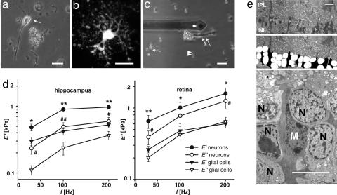
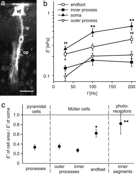
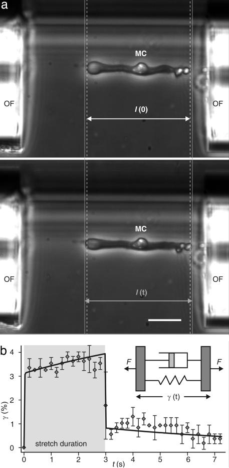
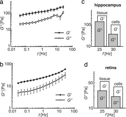
One hundred fifty years ago glial cells were discovered as a second, non-neuronal, cell type in the central nervous system. To ascribe a function to these new, enigmatic cells, it was suggested that they either glue the neurons together (the Greek word "gammalambdaiotaalpha" means "glue") or provide a robust scaffold for them ("support cells"). Although both speculations are still widely accepted, they would actually require quite different mechanical cell properties, and neither one has ever been confirmed experimentally. We investigated the biomechanics of CNS tissue and acutely isolated individual neurons and glial cells from mammalian brain (hippocampus) and retina. Scanning force microscopy, bulk rheology, and optically induced deformation were used to determine their viscoelastic characteristics. We found that (i) in all CNS cells the elastic behavior dominates over the viscous behavior, (ii) in distinct cell compartments, such as soma and cell processes, the mechanical properties differ, most likely because of the unequal local distribution of cell organelles, (iii) in comparison to most other eukaryotic cells, both neurons and glial cells are very soft ("rubber elastic"), and (iv) intriguingly, glial cells are even softer than their neighboring neurons. Our results indicate that glial cells can neither serve as structural support cells (as they are too soft) nor as glue (because restoring forces are dominant) for neurons. Nevertheless, from a structural perspective they might act as soft, compliant embedding for neurons, protecting them in case of mechanical trauma, and also as a soft substrate required for neurite growth and facilitating neuronal plasticity.


Conflict of interest statement

The authors declare no conflict of interest.

Figures

Fig. 1.
Fig. 1.
Comparison of viscoelastic properties of neurons and glial cells. (a–c) Acutely dissociated cells from hippocampus (a, arrow, pyramidal neuron; b, GFAP-EGFP-fluorescent astrocyte) and retina (c, single arrow, bipolar neuron; double arrow, Müller glial cell; double arrowhead, photoreceptor cell) are shown. The tip of the cantilever is also visible in c (single arrowhead). (a and c) Phase-contrast images. (b) Fluorescence image. (d) Elastic storage (E′) and viscous loss modulus (E″) of somata of hippocampal (n = 43) and retinal (bipolar cells, n = 6; amacrine cells, n = 5) neurons and glial cells (hippocampal astrocytes, n = 40; retinal Müller cells, n = 40) (mean ± SEM) is shown. In both types of CNS tissues, glial cells are softer than neurons because Eglia is statistically significant smaller than Eneuron (∗, P < 0.05; ∗∗, P < 0.01, Eneurons vs. E′glia; #, P < 0.05; ##, P < 0.01, Eneurons vs. Eglia), and for all cell types E′/E″ > 1, which means that they have an allover elastically restoring force. (e) (Top) Transmission electron micrograph of the inner nuclear layer (INL) of a guinea pig retina, close to the optic disk. Asterisks indicate Müller cell somata. (Middle) The same image; in the INL, neuronal cell somata are labeled in white, Müller cell somata are in black. Note the more irregular outline of the latter. (Bottom) The image at higher magnification. IPL, inner plexiform layer; M, Müller cell nucleus; N, nuclei of retinal interneurons. (Scale bars: a–c, 20 μm; e, 10 μm.)
Fig. 2.
Fig. 2.
Spatial variation of the cells' mechanical properties. (a) Fluorescence microphotograph of a dye-injected Müller cell (Lucifer Yellow) in a guinea pig retinal slice preparation. ef, endfoot; ip, inner process; s, soma; op, outer process of the Müller cell. (Scale bar: 20 μm.) (b) Comparison of the stiffness of different parts of the radial glial (Müller) cells of the retina (#, P < 0.05; ##, P < 0.01 vs. processes; ∗∗, P < 0.01 vs. all other parts of Müller cells; mean ± SEM). Both cell processes were significantly softer than somata and endfeet, which might be attributed to a different distribution of cell organelles rather than of cytoskeletal elements. (c) Relative stiffness of the respective cellular areas compared with the soma of the according cell (Ecell area/Esoma) at 200 Hz (∗, P < 0.05; ∗∗, P < 0.01 vs. cell processes; mean ± SEM). Processes of Müller cells and pyramidal cells displayed a similar relative stiffness. The endfeet of Müller cells and the inner segments of photoreceptor cells were stiffer with respect to the other cellular compartments, but softer than their somata. Similar relations were observed at 30 and 100 Hz.
Fig. 3.
Fig. 3.
Mechanical deformation and relaxation behavior of a whole Müller (glial) cell (MC) deformed with an optical stretcher. Cells with a length l(0) are aligned with the forces induced on their surface by two infrared laser beams emanating from the optical fibers (OF), whose ends are visible on both sides of the image. (a Upper) Increasing the laser power leads to a stretching of the cells along the laser beam axes. (a Lower) That process results in a cell length l(t). The gray lines help to compare the length before and during the stretching. (Scale bar: 25 μm.) (b) Axial cell strain γ = [I(t) − I(0)]/I(0) as a function of time (mean ± SD). The viscoelastic response of a stretched cell can be modeled by two viscoelastic Voigt elements in series (solid line), resembling a shock absorber. The gray area indicates the duration of the stretching. (Inset) The Voigt model considers a material to be composed of a spring and a dashpot connected in parallel, which represent its elastic and viscous components, respectively. F, forces acting on the viscoelastic object.
Fig. 4.
Fig. 4.
Bulk rheology on CNS tissue. (a and b) Measurements of the complex frequency-dependent shear modulus G* = G′ + iG″ of bovine hippocampal brain slices (n = 9) (a) and retinae (n = 13) (b) (mean ± SEM). Note that the storage modulus G′ always exceeds the loss modulus G″ meaning that both tissues are elastic rather than viscous. (c and d) Comparison between the viscoelastic properties of whole tissues and individual glial cells. There is a good agreement between the data obtained for tissue samples and those calculated for isolated cells [G* = E*/2(l + ν)] even though different animal species were used for methodological reasons.

Similar articles

Cited by

References

    1. Virchow R. Gesammelte Abhandlungen zur Wissenschaftlichen Medicin. Frankfurt, Germany: von Meidinger Sohn; 1856.
    1. Schultze M. Zur Anatomie und Physiologie der Retina. Sohn, Bonn, Germany: von Max Cohen; 1866.
    1. Flanagan LA, Ju YE, Marg B, Osterfield M, Janmey PA. NeuroReport. 2002;13:2411–2415. - PMC - PubMed
    1. Georges PC, Miller WJ, Meaney DF, Sawyer E, Janmey PA. Biophys J. 2006;90:3012–3018. - PMC - PubMed
    1. Arbogast KB, Margulies SS. J Biomech. 1998;31:801–807. - PubMed

Publication types