. 2007 Jan;18(1):229-41.
doi: 10.1091/mbc.e06-06-0570. Epub 2006 Nov 8.

PGY repeats and N-glycans govern the trafficking of paranodin and its selective association with contactin and neurofascin-155

Affiliations

PGY repeats and N-glycans govern the trafficking of paranodin and its selective association with contactin and neurofascin-155

Carine Bonnon et al. Mol Biol Cell. 2007 Jan.

Abstract

Formation of nodes of Ranvier requires contact of axons with myelinating glial cells, generating specialized axo-glial subdomains. Caspr/paranodin is required for the formation of septate-like junctions at paranodes, whereas the related caspr2 is essential for the organization of juxtaparanodes. The molecular mechanisms underlying the segregation of these related glycoproteins within distinct complexes are poorly understood. Exit of paranodin from the endoplasmic reticulum (ER) is mediated by its interaction with F3/contactin. Using domain swapping with caspr2, we mapped a motif with Pro-Gly-Tyr repeats (PGY) in the ectodomain of paranodin responsible for its ER retention. Deletion of PGY allows cell surface delivery of paranodin bypassing the calnexin-calreticulin quality control. Conversely, insertion of PGY in caspr2 or NrCAM blocks these proteins in the ER. PGY is a novel type of processing signal that compels chaperoning of paranodin by contactin. Contactin associated with paranodin is expressed at the cell surface with high-mannose N-glycans. Using mutant CHO lines altered in the processing of N-linked carbohydrates, we show that the high-mannose glycoform of contactin strongly binds neurofascin-155, its glial partner at paranodes. Thus, the unconventional processing of paranodin and contactin may determine the selective association of axo-glial complexes at paranodes.


Figures

Figure 1.
Figure 1.
Schematic representation of paranodin/caspr2 chimeras and mutant constructs. (A) Modular organization of paranodin (pnd), caspr2, and mutated constructs. PndΔPGY1 is deleted of PGY and the beginning of the fourth laminin G (LNG-4) domain (aa 1031–1097). PndΔPGY2 is deleted of part of the second EGF (EGF-2) domain, adjacent interdomain, and PGY motif (aa 985–1081). PndΔ985–1030 is deleted of part of EGF-2 and adjacent interdomain (aa 985-1030). In the caspr2N-pnd chimera, the N-terminal part of paranodin including the discoidin and LNG-1 domains (aa 1–275) is replaced by the corresponding domains of caspr2. The pnd-caspr2C chimera is composed of the N-terminal region of paranodin up to half of the EGF-2 domain (aa 1–984) fused with the C-terminal domain of caspr2 from the LNG-4 domain (aa 1029–1331). The caspr2-PGY construct contains an insertion of a paranodin sequence including half of the EGF-2 domain, PGY, and the beginning of the LNG-4 domain (aa 992–1116) into caspr2. The NrCAM-PGY construct contains the signal peptide and the 6 Ig domains of NrCAM, the GFP and the PGY motif (aa 1014–1083) fused with the C-terminal region of NrCAM from the fourth fibronectin type III up to the transmembrane domain. (B) Putative N-glycosylation sites are indicated along the sequences of paranodin (15 sites) and caspr2 (10 sites). Note that only two of these sites (•) are conserved in the two proteins.
Figure 2.
Figure 2.
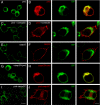
Mapping the ER retention motif in the extracellular region of paranodin. N2a cells were transiently transfected with paranodin (pnd; A and B), paranodin and contactin (C and D), caspr2 (E and F), caspr2N-pnd (G and H), or pnd-caspr2C (I and J) and fixed 48 h after transfection. (A, C, E, G, and I). Cells fixed with methanol were immunostained using anti-paranodin or anti-caspr2 antiserum (green). (B, D, F, H, and J) Cells fixed with paraformaldehyde and permeabilized with TX-100 were double-immunostained using anti-paranodin or anti-caspr2 antiserum (red) and for the ER marker BiP (green). When expressed alone, paranodin is retained in the ER, colocalized with BiP (B). In contrast, when cotransfected with contactin, paranodin is enriched at the cell membrane (C and D). Caspr2 is detected at the cell membrane (E and F). Caspr2N-pnd is only detected in the ER, colocalized with BiP (H). Pnd-caspr2C is mainly expressed at the cell membrane but is also slightly detected in the ER (I and J). Images were obtained using a Leica confocal microscope with 63×/1.3NP lens. A representative image has been selected from the z-stack using a 2× zoom in A, C, E, G, and I (bar, 30 μm) and 2.8× zoom in B, D, F, H, and J (bar, 10 μm).
Figure 3.
Figure 3.
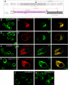
A motif with Pro-Gly-Tyr repeats (PGY) is implicated in the ER retention of paranodin. (A) Sequence alignment of the C-terminal region of the ectodomain of the rat paranodin (ratpnd) and human caspr2 (hucaspr2). A sequence that contains 10 imperfect repeats of Pro-Gly-Tyr (pink) is present between the EGF-2 (gray) and LNG-4 (dark gray) domains of paranodin but not of caspr2. The region deleted in pndΔPGY1 is indicated with a red dashed line and the one deleted in pndΔPGY2 is indicated with a black dashed line. (B–I) Confocal analysis of N2a cells transfected with pndΔPGY1 (B and C), pndΔPGY2 (D and E), pndΔ985–1030 (F and G), or caspr2-PGY (H and I) and fixed 48 h after transfection. (B, D, F, and H) Cells fixed with methanol were immunostained using anti-paranodin or anti-caspr2 antiserum (green). (C, E, G, and I) Cells fixed with paraformaldehyde and permeabilized with TX-100 were double-immunostained using anti-paranodin or anti-caspr2 (red) and anti-BiP (green) antiserum. PndΔPGY1 is retained in the ER (B and C). A different deletion including a sequence upstream of PGY results in the cell membrane expression of pndΔPGY2 (D and E). Deletion of PGY is required for ER exit because in pndΔ985–1030, a shorter deletion restricted to the sequence upstream of PGY does not prevent ER retention (F and G). Reciprocally, insertion of the PGY repeats in caspr2, which normally traffics to the cell surface, induces the ER retention of the caspr2-PGY chimera (H and I). (J and K) N2a cells transfected with NrCAM-GFP and NrCAM-PGY. Insertion of PGY in the extracellular region of NrCAM-GFP results in ER retention of NrCAM-PGY (K), whereas the NrCAM-GFP control construct is only detected at the cell membrane (J). Cells were fixed with paraformaldehyde and confocal images of the GFP fluorescence were collected. Bar, 30 μm in B, D, F, H, J, and K and 10 μm in C, E, G, and I.
Figure 4.
Figure 4.
Role of the PGY repeats analyzed using cell surface biotinylation. Cell surface biotinylation was carried out on N2a cells transfected with paranodin (pnd), pndΔPGY1, pndΔ985–1030, pndΔPGY2, caspr2, caspr2-PGY, or pnd-caspr2C as indicated. Paranodin, pndΔPGY1, pndΔ985–1030, and pndΔPGY2 were immunoprecipitated with an anti-paranodin antibody (lanes 1–4). Caspr2, caspr2-PGY, and pnd-caspr2C were immunoprecipitated with an anti-caspr2 antibody (lanes 5–7). Proteins were revealed with anti-paranodin or anti-caspr2 antiserum, or with peroxidase-conjugated streptavidin. Paranodin, pndΔPGY1, and pndΔ985–1030 are slightly biotinylated (lane 1–3). In contrast, pnd-caspr2C (lane 7) and pndΔPGY2 (lane 4) are strongly biotinylated in the immune precipitate, indicating that they are expressed at the plasma membrane. Biotinylated caspr2 is strongly detected using streptavidin (lane 5), whereas the biotinylated form is dramatically decreased for caspr2-PGY (lane 6), indicating that insertion of PGY inhibits the cell surface expression of the chimera. Ip, immunoprecipitation; Wb, Western blot.
Figure 5.
Figure 5.
Quality control by the calnexin/calreticulin cycle and role of N-glycosylation in the cell membrane delivery of paranodin. N2a cells were transfected with paranodin (pnd) and contactin (A–C), caspr2 (D–F), pndΔPGY2 (G–I), or pnd-caspr2C (J–L). Cells were treated 24 h after transfection with 1 mM castanospermine (B, E, H, and K) or 2 μg/ml tunicamycin (C, F, I, and L) for an additional 18-h period. In double-transfected N2a cells, paranodin is retained in the ER in presence of castanospermine (B), which inhibits the calnexin/calreticulin cycle. Castanospermine treatment does not prevent cell membrane expression of caspr2 (E). Likewise, pndΔPGY2 (H) and pnd-caspr2C (K) expression at the cell membrane is insensitive to castanospermine. Tunicamycin treatment, which blocks the first step of N-glycosylation, results in ER retention of paranodin in double-transfected N2a cells (C) and has no effect on the cell membrane expression of caspr2 (F). Both pndΔPGY2 (I) and pnd-caspr2C (L) are blocked in the ER after tunicamycin treatment. Bar, 10 μm. (M) COS-7 cells were transfected with paranodin, caspr2, or mutant constructs, as indicated. After cell lysis with NP-40, immunoprecipitation was carried out using anti-paranodin antiserum for paranodin (pnd) and pndΔPGY2 (lanes 1–4) or anti-caspr2 antiserum for caspr2, pnd-caspr2C, and caspr2-PGY (lane 5-10). Immunoblots were revealed using anti-paranodin, anti-caspr2, or anti-calnexin antiserum in the lysates (L) and immunoprecipitates (ip). Calnexin is coimmunoprecipitated with paranodin (lane 2) and with pndΔPGY2 (lane 4). Calnexin does not associate with caspr2 (lane 6), is detected at low levels in the pnd-caspr2C immune precipitate (lane 8), and is not detected in the caspr2-PGY immune precipitate (lane 10).
Figure 6.
Figure 6.
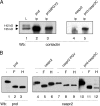
Interaction of paranodin/caspr2 chimeras with contactin and sensitivity to endoglycosidases. (A) Immunoprecipitation from NP-40 lysates of transfected COS-7 cells was carried out using anti-paranodin antiserum for paranodin and pndΔPGY2 (lanes 2 and 3) or anti-caspr2 antiserum for caspr2 and pnd-caspr2C (lanes 4 and 5). Proteins were detected in the lysates (L) and immunoprecipitates (ip) using anti-contactin antiserum. Contactin migrates as a doublet of 135 and 142 kDa in the lysate. Only the 135-kDa form, which is Endo H–sensitive, is detected in the immune precipitate of paranodin, pndΔPGY2, and pnd-caspr2C. As a control, contactin does not coimmunoprecipitate with caspr2. Wb, Western blot. (B) N-glycosylation profiles of paranodin, caspr2, and mutant constructs in transfected COS-7 cells. NP-40 lysates were untreated (−) or incubated with PNGase F (F) or Endo H (H), and immunoblotting was realized with anti-paranodin (lanes 1–3) or anti-caspr2 (lanes 4–12) antiserum. Decreased apparent molecular weight after PNGase F treatment indicates that proteins are N-glycosylated. Paranodin is Endo H–sensitive (lane 3), whereas caspr2 is Endo H–resistant (lane 6). In accordance with its ER distribution, caspr2-PGY is Endo H–sensitive (lane 9). Interestingly, the pnd-caspr2C chimera displays both Endo H–resistant and Endo H–sensitive forms (lane 12).
Figure 7.
Figure 7.
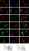
Paranodin requires COPI vesicles for export but does not accumulate in the Golgi after temperature block. (A–C) N2a cells transfected with the dominant negative HA-tagged Arf1(Q71L) were fixed 14 h after transfection. (A) Paranodin coexpressed with contactin (red) is recruited with Arf1(Q71L) (blue) in the Golgi (arrowheads). (B) In cells only transfected with Arf1(Q71L), the ER resident chaperone BiP (red) is recruited for a part in the Golgi with Arf1(Q71L) (green), due to inhibition of the COPI-mediated retrieval from the Golgi to the ER. (C) In contrast, paranodin expressed alone (red) is retained in the ER of cells cotransfected with Arf1(Q71L) (green), indicating that its retention does not result from Golgi-to-ER retrieval. (D–F) N2a cells transfected with contactin-GFP (D) paranodin and contactin-GFP (E), or pnd-caspr2C (F) were incubated during 4 h at 25°C before fixation with methanol 14 h after transfection. After temperature block, contactin-GFP (D, green) strongly accumulates in the Golgi apparatus and colocalizes with the 58K Golgi marker in red (arrowheads). In contrast, in N2a cells coexpressing contactin-GFP (green) and paranodin (red; indicated by asterisks in E) both molecules are distributed in the ER or at the cell membrane but do not colocalize with the 58K Golgi marker in blue (E). Contactin-GFP is concentrated in the Golgi apparatus (arrowheads in E) in a cell that does not coexpress paranodin. Pnd-caspr2C (F, green) is detected in the Golgi apparatus of some of the transfected N2a cells after cold block and colocalized with the 58K Golgi marker in red. Bar, 30 μm. (G) Quantitative analyses of the results presented in A. The percentage of cells with plasma membrane expression of paranodin coexpressed with contactin is significantly reduced by coexpression of Arf1(Q71L) when compared with control conditions (ANOVA, p < 0.01). (H) Quantitative analyses of the results presented in E. Cells were incubated at 37°C for 10 h after transfection (T) and were then incubated for an additional 4-h period at 37°C (control conditions) or were shifted to 25°C. The percentage of cells with plasma membrane expression of paranodin is significantly reduced after incubation at 25°C when compared with control conditions (ANOVA, p < 0.01). Means ± SEM of three independent experiments. More than 100 cells were analyzed in each experiment.
Figure 8.
Figure 8.
NF155-Fc binds contactin bearing high-mannose N-glycans. (A–D) N2a cells transfected with contactin were untreated (A and C) or incubated during 18 h with tunicamycin (B and D). Cells were incubated with NF155-Fc (A and B) or NrCAM-Fc (C and D) at an estimated concentration of 20 μg/ml during 30 min and immunostained using anti-Human Fc antibodies. Tunicamycin treatment prevented NF155-Fc binding. Bar, 40 μm. (E) N2a cells transfected with contactin and paranodin were incubated with NF155-Fc and anti-Human Fc antibodies, fixed, and permeabilized before immunostaining for paranodin. Note that NF155-Fc binding sites colocalize with paranodin. Bar, 20 μm. (F) Schematic representation of the different N-glycan structures anchored at Asn (N) residues in the polypeptide chains and produced by the parental and mutant CHO cell lines. N-acetylglucosamine is abbreviated Gluc-Nac in the legend. (G) Contactin expressed in transfected parental and glycosylation mutant CHO cells was analyzed by SDS-PAGE and immunoblotting. In parental CHO cells, contactin migrates as a doublet of 142 and 135 kDa. By comparison, a single glycosylated variant of contactin is observed in each of the Lec10, Lec1, and Lec23 mutant lines with different apparent molecular weights. An unspecific band indicated with an asterisk at 120 kDa is also detected in untransfected CHO cells. (H) Parental CHO cells and the Lec10, Lec1, and Lec23 mutant lines were transfected with contactin. Immunostaining for contactin on live cells indicates that the different glycoforms of contactin are expressed at the cell surface. (I) Parental CHO cells were transfected with contactin (CHO) or cotransfected with contactin and paranodin (CHO + pnd). Lec10, Lec1, and Lec23 mutant lines were transfected with contactin. Cells were incubated with NF155-Fc at an estimated concentration of 10 μg/ml during 30 min and immunostained using Texas red–conjugated anti-Fc antibodies. All images were collected using identical confocal settings. Bar, 20 μm. (J) Quantitative analysis of the results presented in I. NF155-Fc binding is expressed in arbitrary units corresponding to the mean of fluorescence per cell. More than 40 cells were analyzed in each experiment. NF155-Fc binding significantly increased in the Lec1 and lec23 mutants when compared with parental CHO cells. Similarly, NF155-Fc binding significantly increased in CHO cells cotransfected with paranodin when compared with CHO cells expressing contactin alone (ANOVA, p < 0.01). (K) CHO cells cotransfected with paranodin and contactin were lysed with 1% NP-40. The lysate was untreated (−; lane 1), or incubated with Endo H (H; lane 2), and immunoblotting was performed with both anti-paranodin and anti-contactin antisera. Paranodin (asterisk) detected at 180 kDa is Endo H–sensitive in the lysate. The 142-kDa glycoform of contactin (○) is Endo H–resistant, whereas the135 kDa glycoform of contactin (•) is Endo H–sensitive in the lysate. The lysate was incubated with NF155-Fc linked to protein A-Sepharose and, after washing, bound proteins were eluted and incubated with Endo H (H; lane 3). Paranodin (asterisk) and contactin (•), eluted from the NF155-Fc protein A-Sepharose, are Endo H–sensitive.
Figure 9.
Figure 9.
Model of cooperative interaction between contactin and paranodin. (A) The inset shows the 3-D structure of the PGY motif analyzed by the Rosetta prediction method. The PGY motif (aa1027–1084) is organized with four β-sheets. The PGY motif might display a dynamic structure when paranodin is expressed alone and blocked in the ER. Association with contactin might stabilize the PGY-rich sequence into an organized β-sheet structure, allowing export of paranodin to the plasma membrane (PM). (B) The presence of PGY repeats governs an unconventional processing of paranodin and contactin, which are expressed at the cell surface with ER-type mannose-rich N-glycans. The high-mannose glycoform of contactin displays strong binding activity for NF155, allowing the formation of axo-glial adhesive contacts at paranodes. In contrast, the contactin glycoform expressed at the node, contains complex N-glycans and thereby displays low affinity for NF155.

Similar articles

Cited by

References

    1. Baldwin T. A., Ostergaard H. L. The protein-tyrosine phosphatase CD45 reaches the cell surface via golgi-dependent and -independent pathways. J. Biol. Chem. 2002;277:50333–50340. - PubMed
    1. Bhat M. A., et al. Axon-glia interactions and the domain organization of myelinated axons requires neurexin IV/Caspr/Paranodin. Neuron. 2001;30:369–383. - PubMed
    1. Bichet D., Cornet V., Geib S., Carlier E., Volsen S., Hoshi T., Mori Y., De Waard M. The I-II loop of the Ca2+ channel alpha1 subunit contains an endoplasmic reticulum retention signal antagonized by the beta subunit. Neuron. 2000;25:177–190. - PubMed
    1. Bonnon C., Goutebroze L., Denisenko-Nehrbass N., Girault J. A., Faivre-Sarrailh C. The paranodal complex of F3/contactin and caspr/paranodin traffics to the cell surface via a non-conventional pathway. J. Biol. Chem. 2003;278:48339–48347. - PubMed
    1. Boyle M. E., Berglund E. O., Murai K. K., Weber L., Peles E., Ranscht B. Contactin orchestrates assembly of the septate-like junctions at the paranode in myelinated peripheral nerve. Neuron. 2001;30:385–397. - PubMed

Publication types

MeSH terms