. 2007 Jan;18(1):295-312.
doi: 10.1091/mbc.e06-05-0461. Epub 2006 Nov 8.

Endocytic recycling in yeast is regulated by putative phospholipid translocases and the Ypt31p/32p-Rcy1p pathway

Affiliations

Endocytic recycling in yeast is regulated by putative phospholipid translocases and the Ypt31p/32p-Rcy1p pathway

Nobumichi Furuta et al. Mol Biol Cell. 2007 Jan.

Abstract

Phospholipid translocases (PLTs) have been implicated in the generation of phospholipid asymmetry in membrane bilayers. In budding yeast, putative PLTs are encoded by the DRS2 gene family of type 4 P-type ATPases. The homologous proteins Cdc50p, Lem3p, and Crf1p are potential noncatalytic subunits of Drs2p, Dnf1p and Dnf2p, and Dnf3p, respectively; these putative heteromeric PLTs share an essential function for cell growth. We constructed temperature-sensitive mutants of CDC50 in the lem3Delta crf1Delta background (cdc50-ts mutants). Screening for multicopy suppressors of cdc50-ts identified YPT31/32, two genes that encode Rab family small GTPases that are involved in both the exocytic and endocytic recycling pathways. The cdc50-ts mutants did not exhibit major defects in the exocytic pathways, but they did exhibit those in endocytic recycling; large membranous structures containing the vesicle-soluble N-ethylmaleimide-sensitive factor attachment protein receptor Snc1p intracellularly accumulated in these mutants. Genetic results suggested that the YPT31/32 effector RCY1 and CDC50 function in the same signaling pathway, and simultaneous overexpression of CDC50, DRS2, and GFP-SNC1 restored growth as well as the plasma membrane localization of GFP-Snc1p in the rcy1Delta mutant. In addition, Rcy1p coimmunoprecipitated with Cdc50p-Drs2p. We propose that the Ypt31p/32p-Rcy1p pathway regulates putative phospholipid translocases to promote formation of vesicles destined for the trans-Golgi network from early endosomes.


Figures

Figure 1.
Figure 1.
Cdc50 family proteins form complexes with Drs2 family proteins. (A) Coimmunoprecipitation of Lem3p and Crf1p with Dnf2p and Dnf3p, respectively. Cells were grown at 25°C to a cell density of 0.5 OD600/ml in YPDA medium. Membrane extracts were then prepared as described in Materials and Methods. Myc-tagged Dnf2p or Dnf3p was immunoprecipitated with an anti-Myc antibody from these extracts. Immunoprecipitates were subjected to SDS-PAGE, followed by immunoblot analysis using antibodies against Lem3p or HA (top) and Myc (bottom). The results shown are representative of several experiments. The strains used were as follows: YKT1099 (DNF2-Myc LEM3) and YKT1098 (DNF2 LEM3) (left) and YKT1100 (DNF3-Myc CRF1-HA) and YKT1098 (DNF3 CRF1-HA) (right). (B) Localization of Dnf2p-EGFP. Wild-type (YKT921) and lem3Δ (YKT923) cells containing the DNF2-EGFP construct in the genome were grown to early to mid-logarithmic phase in YPDA medium at 30°C and immediately observed by fluorescence microscopy. (C) Localization of Dnf3p-EGFP. Wild-type (YKT925) and crf1Δ (YKT928) cells containing the DNF3-EGFP construct in the genome were grown and observed as described in (B). Bars, 5 μm.
Figure 2.
Figure 2.
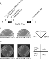
Overexpression of YPT32 or YPT31 suppresses the temperature-sensitive growth defect of the cdc50-ts mutants. (A) The domain structure of Cdc50p and the amino acid substitutions in Cdc50-11p and Cdc50-162p. The black boxes indicate potential transmembrane domains. (B) Suppression of the cdc50-ts mutations by multicopy YPT32 or YPT31. cdc50-11 (YKT993) and cdc50-162 (YKT942) cells were transformed with pKT1555 (YEplac181-YPT32), pKT1554 (YEplac181-YPT31), pKT1259 (YEplac181-CDC50), or a control vector (YEplac181). Transformants were streaked onto a YPDA plate, followed by incubation at 37°C for 2 d. (C) The GTP-bound form, but not the GDP-bound form, of Ypt32p suppresses the ts growth defect of cdc50-ts mutants. cdc50-11 (YKT993) and cdc50-162 (YKT942) mutant cells were transformed with pKT1578 (YEplac181-YPT32Q72L), pKT1579 (YEplac181-YPT32S27N), pKT1555 (YEplac181-YPT32), or a control vector (YEplac181). For each plasmid, four independent transformants were streaked onto a YPDA plate, followed by incubation at 37°C for 3 d.
Figure 3.
Figure 3.
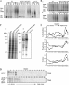
Secretion and formation of secretory vesicles are nearly normal in a cdc50-ts mutant. (A and B) Pulse-chase experiments of invertase secretion. Wild-type (YKT38), cdc50-11 (YKT993), and sec2-56 (ANS2-3A) cells (A) or wild-type (YKT38) and cdc50-11 (YKT993) cells (B) were grown for 2.5 h at 37°C, induced to produce invertase for 30 min, pulse labeled with Tran35S-label for 7 min (A) or 2 min (B), and chased for the indicated time at 37°C. Samples were separated by centrifugation into internal (I) and external (E) fractions. Invertase was recovered by immunoprecipitation and visualized by SDS-PAGE and a phosphorimager system. (C) General secretion by the cdc50-11 mutant. Wild-type (YKT38), cdc50-11 (YKT993), and sec2-56 (ANS2-3A) cells were grown for 2.5 h at 37°C, pulse labeled with Tran35S-label for 15 min, and chased for 45 min. Samples were separated by centrifugation into I and E fractions. Proteins in each fraction were precipitated with TCA and visualized by SDS-PAGE and a phosphorimager system. (D) Western blots for Pma1p, a marker for low-density vesicles, in Nycodenz gradient fractions. sec6-4 (YKT1010) and cdc50-11 sec6-4 (YKT1011) cells transformed with pKT1486 (PACT1-SUC2) were incubated for 2 h at 37°C. Secretory vesicles were prepared and fractionated as described in Materials and Methods. Numbered fractions (as indicated at the top) were heated in sample buffer for 15 min at 37°C, separated by SDS-PAGE, and probed with antibodies against Pma1p. (E) Activities of marker enzymes for high-density vesicles in Nycodenz gradient fractions. Fractions were prepared from sec6-4 (diamonds) and cdc50-11 sec6-4 (triangles) cells as described in D. Hydrolyzing enzyme activities are expressed in arbitrary units based on the absorbance measured at 415 nm (acid phosphatase, top; exoglucanase, middle) or at 540 nm (invertase, bottom).
Figure 4.
Figure 4.
Normal endocytic and VPS pathways in the cdc50-ts mutants. (A) Internalization and transport of FM4-64 to the vacuole. Wild-type (YKT38), cdc50-11 (YKT993), and cdc50-162 (YKT942) cells grown for 3 h were stained with FM4-64 for 15 min in YPDA medium, and chased for 30 min in fresh YPDA medium, at the indicated temperature. Bar, 5 μm. (B) Pulse-chase experiments of CPY intracellular transport. Wild-type (YKT38) and cdc50-11 (YKT993) cells were grown for 2.5 h at 37°C, pulse labeled with Tran35S-label for 15 min, and chased for 45 min at 37°C. CPY was immunoprecipitated from I and E fractions, resolved by SDS-PAGE, and visualized using a phosphorimager system. (C) Secretion of CPY. Wild-type (YKT38), cdc50-11 (YKT993), vps1Δ (KKT276), vps4Δ (KKT277), and vps30Δ (AKY15) cells were grown for 3 h at 37°C in contact with a nitrocellulose filter, and secreted CPY was detected by probing with antibodies against CPY. (D) Localization of Vps10p-EGFP. Wild-type (YKT957), cdc50- 11 (YKT1086), and cdc50-162 (YKT1088) cells containing the VPS10-EGFP construct in the genome were grown for 3 h at 25 or 37°C in SD medium. Bar, 5 μm. (E) Localization of GFP-Pep12p. Wild-type (YKT38), cdc50-11 (YKT993), and cdc50-162 (YKT942) cells transformed with pKT1487 (pRS416-GFP-PEP12) were grown for 3 h at 25°C or 37°C in SDA-Ura medium. Bar, 5 μm.
Figure 5.
Figure 5.
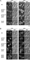
Recycling marker proteins are mislocalized in the cdc50-ts mutants. (A) Localization of Kex2p-EGFP. Wild-type (YKT903), cdc50-11 (YKT1000), cdc50-162 (YKT1001), and lem3Δ crf1Δ (YKT1310) cells containing the KEX2-EGFP construct in the genome were grown for 3 h at 25 or 37°C in SD medium. (B) Localization of GFP-Snc1p. Wild-type (YNF63), cdc50-11 (YNF65), cdc50-162 (YNF67), lem3Δ crf1Δ (YNF61), and dnf1Δ dnf2Δ dnf3Δ (YNF784) cells carrying the pRS416-GFP-SNC1 plasmid were grown for 3 h at 25 or 37°C in SDA-Ura medium. Bars, 5 μm.
Figure 6.
Figure 6.
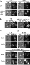
The cdc50-ts mutant is defective in plasma membrane-to-TGN transport. (A) Plasma membrane localization of GFP-Snc1p is restored in the cdc50-ts mutant by blocking endocytosis with LAT-A. Wild-type (YNF63) and cdc50-11 (YNF65) cells carrying the pRS416-GFP-SNC1 plasmid were preincubated at 37°C for 3 h and then treated with 100 μM LAT-A or DMSO (vehicle control) for 10 min at 37°C in SDA-Ura medium. (B) Localization of GFP-Snc1p-pm. Wild-type (YKT38) and cdc50-11 (YKT993) cells transformed with pRS416-GFP-SNC1 pm were grown to early logarithmic phase at 25°C and shifted to 37°C for 3 h in SDA-Ura medium. (C) Pretreatment with LAT-A inhibits abnormal accumulation of GFP-Snc1p in the cdc50-ts mutant. Wild-type (YNF63) and cdc50-11 (YNF65) cells carrying the pRS416-GFP-SNC1 plasmid were grown to early logarithmic phase at 25°C and were then pretreated with 100 μM LAT-A or DMSO (vehicle control) for 10 min at 25°C in SDA-Ura medium (0 min), followed by incubation at 37°C for 30 min in SDA-Ura medium in the presence (LAT-A) or absence (DMSO) of 100 μM LAT-A (30 min). Bars, 5 μm.
Figure 7.
Figure 7.
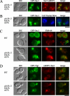
GFP-Snc1p and GFP-Tlg1p accumulate in early endosome-derived structures in the cdc50-ts mutant. (A) GFP-Snc1p and the TGN marker Sec7p-mRFP1 are not colocalized in the cdc50-11 mutant. cdc50-11 SEC7-mRFP1 (YNF153) cells carrying pRS416-GFP-SNC1 were incubated at 37°C for 3 h in SDA-Ura medium. Obtained images were merged to compare the two signal patterns. (B) GFP-Snc1p is not localized to vacuoles stained with CellTracker Blue CMAC in the cdc50-11 mutant. cdc50-11 (YNF65) cells carrying the pRS416-GFP-SNC1 plasmid were grown at 37°C for 3 h, followed by staining with 100 μM CellTracker Blue CMAC at 37°C for 15 min. (C) GFP-Snc1p was colocalized with FM4-64 after a short incubation in the cdc50-11 mutant. Wild-type (YNF63) and cdc50-11 (YNF65) cells carrying the pRS416-GFP-SNC1 plasmid were grown at 37°C for 3 h, stained with 32 μM FM4-64 on ice for 30 min, and chased in fresh medium at 37°C for 10 min. (D) mRFP1-Snc1p and GFP-Tlg1p are colocalized in the cdc50-11 mutant. Wild-type (YKT38) and cdc50-11 (YKT993) cells cotransformed with pKT1566 (YEplac181-GFP-TLG1) and pKT1563 (pRS416-mRFP1-SNC1) were incubated at 37°C for 3 h in SD-Leu-Ura medium, followed by microscopic examination after fixation with 0.5% formaldehyde. Bars, 5 μm.
Figure 8.
Figure 8.
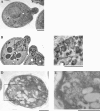
Electron microscopic examination of the abnormal structures containing HA-Snc1p in the cdc50-ts mutant. (A–C) The electron microscopic observation was performed using the glutaraldehyde-permanganate fixation technique. Wild-type (YKT38; A) and cdc50-11 mutant cells (YKT993; B and C) were grown at 25°C to early logarithmic phase, shifted to 37°C, and grown in YPDA medium for 3 h. Cells were prepared for EM as described in Materials and Methods. The boxed region in B is enlarged in C. An arrow and an arrowhead indicate a horseshoe-like structure and a double-membrane ring, respectively. Bars, 1 μm. (D and E) Immunoelectron microscopic observation of the cdc50-11 mutant was performed by the aldehyde fixation/metaperiodate permeabilization method. cdc50-11 mutant cells (YKT993) transformed with pKT1564 (pRS416-HA-SNC1) were grown at 25°C to early logarithmic phase, shifted to 37°C, and grown in SDA-Ura medium for 3 h. Cells were prepared for immuno-EM as described in Materials and Methods and labeled with an anti-HA antibody. The boxed region in D is enlarged in E. Bars, 1 μm.
Figure 9.
Figure 9.
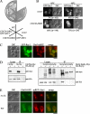
The Cdc50p–Drs2p complex functionally and physically interacts with Rcy1p. (A) Suppression of the growth defect of the rcy1Δ mutant by simultaneous overexpression of CDC50, DRS2, and GFP-SNC1. rcy1Δ mutant cells (YKT951) were cotransformed with combinations of plasmids as follows: pKT1472 (YEplac195-DRS2-CDC50) and pKT1490 (pRS315-GFP-SNC1) for DRS2-CDC50 GFP-SNC1, pKT1472 (YEplac195-DRS2-CDC50) and pRS315 for DRS2-CDC50, YEplac195 and pKT1490 (pRS315-GFP-SNC1) for GFP-SNC1, and YEplac195 and pRS315 for vector. Transformants and wild-type cells (YKT38) were streaked onto a YPDA plate, followed by incubation at 18°C for 9 d. (B) Simultaneous overexpression of CDC50, DRS2, and GFP-SNC1 partially restored the plasma membrane localization of GFP-Snc1p in the rcy1Δ mutant. rcy1Δ mutant cells (YKT951) were cotransformed with pKT1490 (pRS315-GFP-SNC1) and a control vector (YEplac195; top) or pKT1472 (YEplac195-DRS2-CDC50; bottom). Cells were incubated at 18 or 30°C for 12 h in SD-Leu-Ura medium. Numbers indicate the percentages of cells in which GFP-Snc1p was localized to the plasma membrane. Bar, 5 μm. (C) GFP-Rcy1p partially colocalized with Drs2p-mRFP1. DRS2-mRFP1 (YKT871) cells transformed with pKT1560 (YEplac181-GFP-RCY1) were grown to early to mid-logarithmic phase at 30°C in SD-Leu-Ura medium. Obtained images were merged to compare the two signal patterns. Bar, 5 μm. (D) Coimmunoprecipitation of Rcy1p with Cdc50p, Drs2p, Dnf1p, and Dnf2p. Cells grown to mid-logarithmic phase at 30°C in SDA-Ura medium. Membrane extracts were then prepared as described in Materials and Methods. Myc-tagged Cdc50p, Drs2p, Dnf1p, or Dnf2p were immunoprecipitated with an anti-Myc antibody from membrane extracts. Immunoprecipitates were subjected to SDS-PAGE, followed by immunoblot analysis using antibodies against Myc (top) and HA (bottom). The results shown are representative of several experiments. The yeast strains used were as follows: YKT1101 (CDC50-Myc) and YKT38 (CDC50) (left); YKT792 (DRS2-Myc), YKT760 (DNF1-Myc), YKT1062 (DNF2-Myc), and YKT38 (DRS2 DNF1 DNF2) (right). All these strains carried pKT1626 (YEplac195-HA-BS-RCY1). (E) Colocalization of Cdc50p-EGFP with mRFP1-Snc1p in the rcy1Δ mutant. CDC50-EGFP (YKT259) and rcy1Δ CDC50-EGFP (YKT1102) cells transformed with pKT1563 (pRS416-mRFP1-SNC1) were grown to mid-logarithmic phase at 30°C in SDA-Ura medium. Bar, 5 μm.

Similar articles

Cited by

References

    1. Alder-Baerens N., Lisman Q., Luong L., Pomorski T., Holthuis J. C. Loss of P4 ATPases Drs2p and Dnf3p disrupts aminophospholipid transport and asymmetry in yeast post-Golgi secretory vesicles. Mol. Biol. Cell. 2006;17:1632–1642. - PMC - PubMed
    1. Ayscough K. R., Stryker J., Pokala N., Sanders M., Crews P., Drubin D. G. High rates of actin filament turnover in budding yeast and roles for actin in establishment and maintenance of cell polarity revealed using the actin inhibitor latrunculin-A. J. Cell Biol. 1997;137:399–416. - PMC - PubMed
    1. Bai C., Sen P., Hofmann K., Ma L., Goebl M., Harper J. W., Elledge S. J. SKP1 connects cell cycle regulators to the ubiquitin proteolysis machinery through a novel motif, the F-box. Cell. 1996;86:263–274. - PubMed
    1. Benli M., Doring F., Robinson D. G., Yang X., Gallwitz D. Two GTPase isoforms, Ypt31p and Ypt32p, are essential for Golgi function in yeast. EMBO. J. 1996;15:6460–6475. - PMC - PubMed
    1. Black M. W., Pelham H. R. A selective transport route from Golgi to late endosomes that requires the yeast GGA proteins. J. Cell Biol. 2000;151:587–600. - PMC - PubMed

Publication types

MeSH terms