

Cocaine increases the intracellular calcium concentration in brain independently of its cerebrovascular effects
- PMID: 17093073
- PMCID: PMC6674780
- DOI: 10.1523/JNEUROSCI.3612-06.2006
Cocaine increases the intracellular calcium concentration in brain independently of its cerebrovascular effects
Abstract
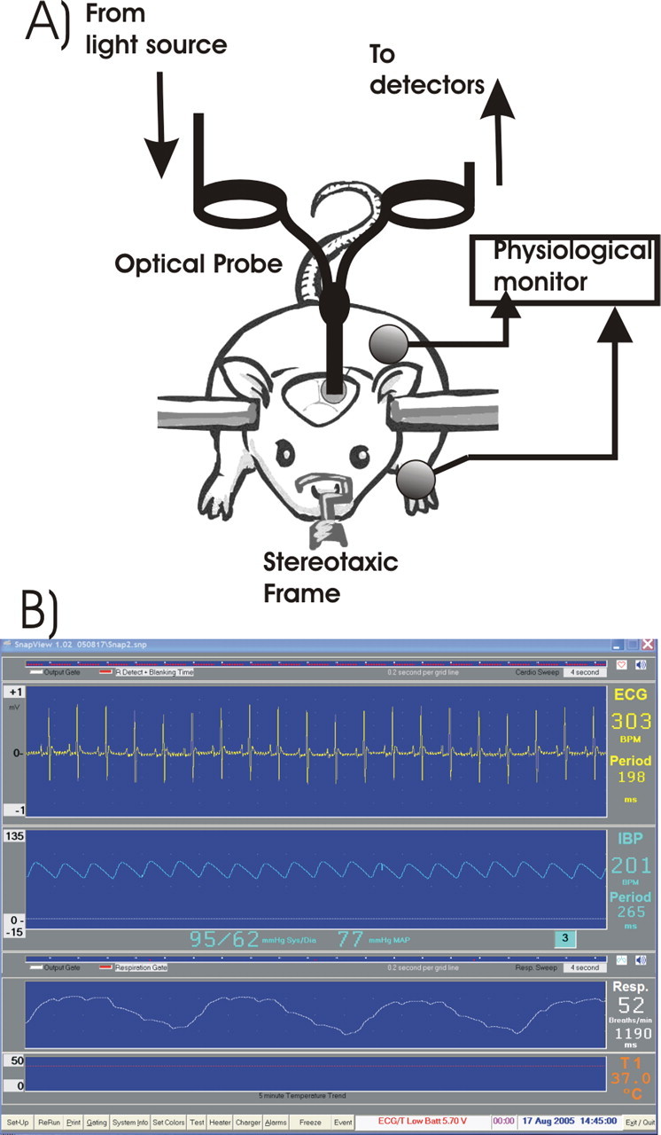
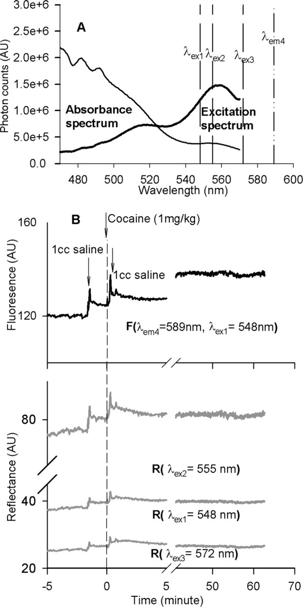
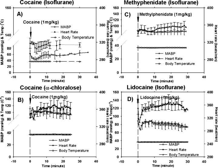
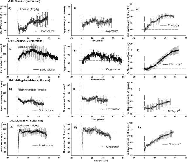
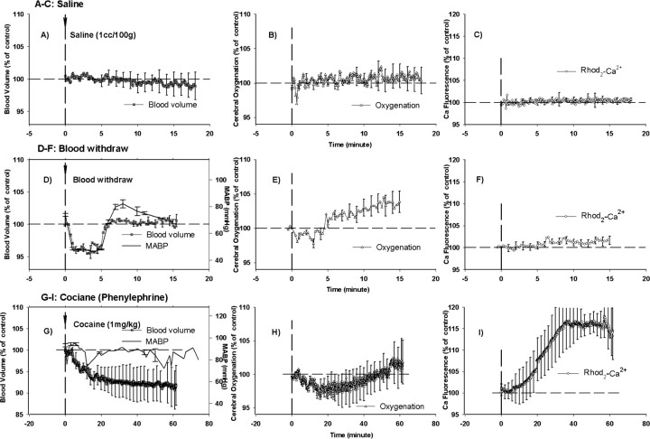
Cocaine abuse increases the risk of life-threatening neurological complications such as strokes and seizures. Although the vasoconstricting properties of cocaine underlie its cerebrovascular effects, the mechanisms underlying its neurotoxicity remain incompletely understood. Here, we use optical techniques to measure cerebral blood volume, hemoglobin oxygenation (S(t)O(2)), and intracellular calcium ([Ca(2+)](i)) to test the hypothesis that cocaine increases [Ca(2+)](i) in the brain. The effects of cocaine were compared with those of methylphenidate, which has similar catecholaminergic effects as cocaine (except for serotonin increases) but no local anesthetic properties, and of lidocaine, which has similar local anesthetic effects as cocaine but is devoid of catecholaminergic actions. To control for the hemodynamic effects of cocaine, we assessed the effects of cocaine in animals in which normal blood pressure was maintained by infusion of phenylephrine, and we also measured the effects of transient hypotension (mimicking that induced by cocaine). We show that cocaine induced significant increases ( approximately 10-15%) in [Ca(2+)](i) that were independent of its hemodynamic effects and of the anesthetic used (isofluorance or alpha-chloralose). Lidocaine but not methylphenidate also induced significant [Ca(2+)](i) increases ( approximately 10-13%). This indicates that cocaine at a dose within the range used by drug users significantly increases the [Ca(2+)](i) in the brain and its local anesthetic, but neither its catecholaminergic nor its hemodynamic actions, underlies this effect. Cocaine-induced [Ca(2+)](i) increases are likely to accentuate the neurotoxic effects from cocaine-induced vasoconstriction and to facilitate the occurrence of seizures from the catecholaminergic effects of cocaine. These findings support the use of calcium channel blockers as a strategy to minimize the neurotoxic effects of cocaine.
Figures
References
-
- Astrup J, Symon L, Branston NM, Lassen NA. Cortical evoked potential and extracellular K+ and H+ at critical levels of brain ischemia. Stroke. 1977;8:51–57. - PubMed
-
- Bartzokis G, Beckson M, Lu PH, Edwards N, Rapoport R, Bridge P, Mintz J. Cortical gray matter volumes are associated with subjective responses to cocaine infusion. Am J Addict. 2004;13:64–73. - PubMed
-
- Benveniste H, Jorgensen MB, Diemer NH, Hansen AJ. Calcium accumulation by glutamate receptor activation is involved in hippocampal cell damage after ischemia. Acta Neurol Scand. 1988;78:529–536. - PubMed
-
- Berwick J, Devonshire IM, Martindale AJ, Johnston D, Zheng Y, Kennerley AJ, Overton PG, Mayhew JE. Cocaine administration produces a protracted decoupling of neural and haemodynamic responses to intense sensory stimuli. Neuroscience. 2005;132:361–374. - PubMed
-
- Bolouri MR, Small GA. Neuroimaging of hypoxia and cocaine-induced hippocampal stroke. J Neuroimaging. 2004;14:290–291. - PubMed
Publication types
MeSH terms
Substances
Grants and funding
LinkOut - more resources
Full Text Sources
Miscellaneous